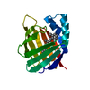
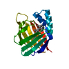
PDB-4ybp: Crystal structure of the R111K:Y134F:T54V:R132Q:P39Q:R59Y mutant of human cellular retinoic acid binding proteinii with retinal after 24 hour incubation at 1.83 angstrom resolution - thermodynamic product - 1st cycle Method: X-RAY DIFFRACTION / Resolution: 1.831 Å
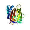
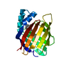
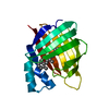
PDB-4ybu: Crystal structure of the R111K:Y134F:T54V:R132Q:P39Q:R59Y mutant of human Cellular Retinoic Acid Binding ProteinII in complex with Retinal after 24 h incubation and 1 hour UV irradiation at 1.92 angstrom - 1st cycle Method: X-RAY DIFFRACTION / Resolution: 1.924 Å
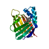
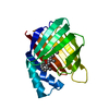
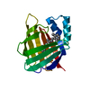
PDB-4yce: Crystal structure of the R111K:Y134F:T54V:R132Q:P39Q:R59Y mutant of human cellular retinoic acid binding proteinii with retinal at 1.95 angstrom- visible light irradiated crystal for 1 hour - 2nd cycle Method: X-RAY DIFFRACTION / Resolution: 1.953 Å
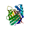
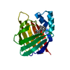
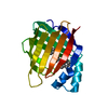
PDB-4ych: Crystal Structure of R111K:Y134F:T54V:R132Q:P39Q:R59Y mutant of human Cellular Retinoic Acid Binding ProteinII with Retinal at 1.96 Angstrom - UV irradiated Crystal for 1 hour - 2nd cycle Method: X-RAY DIFFRACTION / Resolution: 1.96 Å
PDB-4yda: Crystal structure of R111K:Y134F:T54V:R132Q:P39Q:R59Y mutant of human cellular retinoic acid binding proteinii with retinal at 1.95 angstrom - after 1hour visible light irradiation - 3rd cycle Method: X-RAY DIFFRACTION / Resolution: 1.951 Å
PDB-4ydb: Crystal structure of the R111K:Y134F:T54V:R132Q:P39Q:R59Y mutant of human Cellular Retinoic Acid Binding Protein II in complex with Retinal at 2.03 angstrom -UV irradiated crystal- 3rd cycle Method: X-RAY DIFFRACTION / Resolution: 2.03 Å
PDB-4yfp: CRYSTAL STRUCTURE OF THE R111K:Y134F:T54V:R132Q:P39Y:R59Y MUTANT OF HUMAN CELLULAR RETINOIC ACID BINDING PROTEIN II WITH RETINAL AFTER 20 MINUTES INCUBATION AT 1.95 ANGSTROM RESOLUTION-Kinetic Product Method: X-RAY DIFFRACTION / Resolution: 1.951 Å
PDB-4yfq: CRYSTAL STRUCTURE OF THE R111K:Y134F:T54V:R132Q:P39Y:R59Y MUTANT OF HUMAN CELLULAR RETINOIC ACID BINDING PROTEINII WITH RETINAL AFTER 24 HOURS INCUBATION AT 1.62 ANGSTROM RESOLUTION - Thermodynamic product - 1st cycle Method: X-RAY DIFFRACTION / Resolution: 1.62 Å
PDB-4yfr: Crystal structure of the R111K:Y134F:T54V:R132Q:P39Y:R59Y mutant of human Cellular Retinoic Acid Binding Protein II with Retinal at 1.95 Angstrom Resolution - UV irradiated crystal for 30 minutes - 1st Cycle Method: X-RAY DIFFRACTION / Resolution: 1.952 Å
PDB-4ygg: CRYSTAL STRUCTURE OF R111K:Y134F:T54V:R132Q:P39Y:R59Y MUTANT OF HUMAN CELLULAR RETINOIC ACID BINDING PROTEIN II WITH RETINAL AT 1.9 ANGSTROM RESOLUTION - VISIBLE LIGHT IRRADIATED CRYSTAL - 2ND CYCLE Method: X-RAY DIFFRACTION / Resolution: 1.9 Å
PDB-4ygh: Crystal Structure of R111K:Y134F:T54V:R132Q:P39Y:R59Y mutant of human Cellular Retinoic Acid Binding Protein II with Retinal at 2.1 Angstrom resolution - UV irradiated crystal - 2nd cycle Method: X-RAY DIFFRACTION / Resolution: 2.1 Å
PDB-4ygz: CRYSTAL STRUCTURE OF THE R111K:Y134F:T54V:R132Q:P39Y:R59Y MUTANT OF HUMAN CELLULAR RETINOIC ACID BINDING PROTEIN II WITH RETINAL AT 2.06 ANGSTROM RESOLUTION - VISIBLE LIGHT IRRADIATED CRYSTAL -3RD CYCLE Method: X-RAY DIFFRACTION / Resolution: 2.06 Å
PDB-4yh0: Crystal structure of the R111K:Y134F:T54V:R132Q:P39Y:R59Y mutant of human Cellular Retinoic Acid Binding Protein II in complex with Retinal at 2.14 angstrom resolution - UV irradiated crystal - 3rd cycle Method: X-RAY DIFFRACTION / Resolution: 2.144 Å
PDB-4ykm: Crystal structure of the R111K:Y134F:T54V:R132Q:P39Q:R59Y:A32W:F3Q mutant of human Cellular Retinoic Acid Binding Protein II with Retinal at 1.58 angstrom resolution Method: X-RAY DIFFRACTION / Resolution: 1.58 Å
PDB-4yko: Crystal structure of the R111K:Y134F:T54V:R132Q:P39Q:R59Y:A32Y:F3Q mutant of human Cellular Retinoic Acid Binding Protein II with Retinal at 1.58 angstrom resolution Method: X-RAY DIFFRACTION / Resolution: 1.57 Å
In the structure databanks used in Yorodumi, some data are registered as the other names, "COVID-19 virus" and "2019-nCoV". Here are the details of the virus and the list of structure data.
Jan 31, 2019. EMDB accession codes are about to change! (news from PDBe EMDB page)
EMDB accession codes are about to change! (news from PDBe EMDB page)
The allocation of 4 digits for EMDB accession codes will soon come to an end. Whilst these codes will remain in use, new EMDB accession codes will include an additional digit and will expand incrementally as the available range of codes is exhausted. The current 4-digit format prefixed with “EMD-” (i.e. EMD-XXXX) will advance to a 5-digit format (i.e. EMD-XXXXX), and so on. It is currently estimated that the 4-digit codes will be depleted around Spring 2019, at which point the 5-digit format will come into force.
The EM Navigator/Yorodumi systems omit the EMD- prefix.
Related info.:Q: What is EMD? / ID/Accession-code notation in Yorodumi/EM Navigator
 Movie
Movie Controller
 Structure viewers
 About Yorodumi Papers


 Authors
 External links
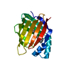


 Keywords
 homo sapiens (human)