 ムービー
ムービー コントローラー


+ データを開く

- 基本情報
基本情報
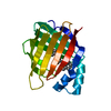
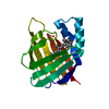
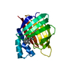
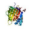
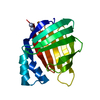
| 登録情報 | データベース: PDB / ID: 6mqw | ||||||
|---|---|---|---|---|---|---|---|
| タイトル | Crystal Structure of All-trans Retinal-Bound R111K:Y134F:T54V:R132Q:P39Y:R59Y:L121E Human Cellular Retinoic Acid Binding Protein II Irradiated with 400 nm laser (30 seconds) and subsequently dark adapted (10 minutes) at 2.1 Angstrom Resolution | ||||||
 要素 要素 | Cellular retinoic acid-binding protein 2 | ||||||
 キーワード キーワード | LIPID BINDING PROTEIN / iLBP / Rhodopsin mimic | ||||||
| 機能・相同性 |  機能・相同性情報 機能・相同性情報positive regulation of collateral sprouting / retinoid binding / retinoic acid binding / retinal binding / embryonic forelimb morphogenesis / retinoic acid metabolic process / retinol binding / Signaling by Retinoic Acid / epidermis development / fatty acid transport ...positive regulation of collateral sprouting / retinoid binding / retinoic acid binding / retinal binding / embryonic forelimb morphogenesis / retinoic acid metabolic process / retinol binding / Signaling by Retinoic Acid / epidermis development / fatty acid transport / cyclin binding / fatty acid binding / regulation of DNA-templated transcription / endoplasmic reticulum / signal transduction / extracellular exosome / nucleoplasm / nucleus / cytoplasm / cytosol 類似検索 - 分子機能 | ||||||
| 生物種 |  Homo sapiens (ヒト) Homo sapiens (ヒト) | ||||||
| 手法 |  X線回折 / シンクロトロン / 分子置換 / 解像度: 2.09 Å X線回折 / シンクロトロン / 分子置換 / 解像度: 2.09 Å | ||||||
 データ登録者 データ登録者 | Ghanbarpour, A. / Geiger, J. | ||||||
| 資金援助 |  米国, 1件 米国, 1件
| ||||||
 引用 引用 | ジャーナル: J. Am. Chem. Soc. / 年: 2019 タイトル: Mimicking Microbial Rhodopsin Isomerization in a Single Crystal. 著者: Ghanbarpour, A. / Nairat, M. / Nosrati, M. / Santos, E.M. / Vasileiou, C. / Dantus, M. / Borhan, B. / Geiger, J.H. | ||||||
| 履歴 |
|
- 構造の表示
構造の表示
| 構造ビューア | 分子: MolmilJmol/JSmol |
|---|
- ダウンロードとリンク
-ダウンロード
| PDBx/mmCIF形式 | 6mqw.cif.gz | 44.9 KB | 表示 | PDBx/mmCIF形式 |
|---|---|---|---|---|
| PDB形式 | pdb6mqw.ent.gz | 30.2 KB | 表示 | PDB形式 |
| PDBx/mmJSON形式 | 6mqw.json.gz | ツリー表示 | PDBx/mmJSON形式 | |
| その他 |  その他のダウンロード その他のダウンロード |
-検証レポート
| アーカイブディレクトリ | https://data.pdbj.org/pub/pdb/validation_reports/mq/6mqwftp://data.pdbj.org/pub/pdb/validation_reports/mq/6mqw | HTTPS FTP |
|---|
-関連構造データ
| 関連構造データ |  6mopC  6moqC  6morC  6movC  6moxC  6mpkC  6mqiC  6mqjC  6mqxC  6mqyC  6mqzC  6mr0C 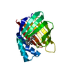 4yfpS S: 精密化の開始モデル C: 同じ文献を引用 ( |
|---|---|
| 類似構造データ |


-リンク
 PDBj
PDBj


- 集合体
集合体
| 登録構造単位 | 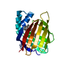
| ||||||||
|---|---|---|---|---|---|---|---|---|---|
| 1 |
| ||||||||
| 2 | 
| ||||||||
| 単位格子 |
|
-要素
| #1: タンパク質 | 分子量: 15594.747 Da / 分子数: 1 / 由来タイプ: 組換発現 / 由来: (組換発現) Homo sapiens (ヒト) / 遺伝子: CRABP2 / 発現宿主:  |
|---|---|
| #2: 化合物 | ChemComp-RET /   分子量: 284.436 Da / 分子数: 1 / 由来タイプ: 合成 / 式: C20H28O 分子量: 284.436 Da / 分子数: 1 / 由来タイプ: 合成 / 式: C20H28O |
| #3: 水 | ChemComp-HOH /  分子量: 18.015 Da / 分子数: 54 / 由来タイプ: 天然 / 式: H2O 分子量: 18.015 Da / 分子数: 54 / 由来タイプ: 天然 / 式: H2O |
| Has protein modification | Y |
-実験情報
-実験
| 実験 | 手法: X線回折 / 使用した結晶の数: 1 |
|---|
- 試料調製
試料調製
| 結晶 | マシュー密度: 3.18 Å3/Da / 溶媒含有率: 61.35 % / 解説: hexagonal |
|---|---|
| 結晶化 | 温度: 277 K / 手法: 蒸気拡散法, ハンギングドロップ法 / pH: 6.5 / 詳細: PEG 3350, DL-malic acid / PH範囲: 6.0-6.5 |
-データ収集
| 回折 | 平均測定温度: 100 K / Serial crystal experiment: N |
|---|---|
| 放射光源 | 由来: シンクロトロン / サイト: APS / ビームライン: 21-ID-D / 波長: 0.97911 Å |
| 検出器 | タイプ: DECTRIS EIGER X 9M / 検出器: PIXEL / 日付: 2016年12月21日 |
| 放射 | プロトコル: SINGLE WAVELENGTH / 単色(M)・ラウエ(L): M / 散乱光タイプ: x-ray |
| 放射波長 | 波長: 0.97911 Å / 相対比: 1 |
| 反射 | 解像度: 2.09→35.645 Å / Num. obs: 12273 / % possible obs: 99.9 % / 冗長度: 9.5 % / Rmerge(I) obs: 0.067 / Rrim(I) all: 0.093 / Net I/σ(I): 36.75 |
| 反射 シェル | 解像度: 2.09→2.12 Å / 冗長度: 9.7 % / Rmerge(I) obs: 0.677 / Mean I/σ(I) obs: 3.8 / Num. unique obs: 1205 / Rrim(I) all: 0.754 / % possible all: 100 |
- 解析
解析
| ソフトウェア |
| |||||||||||||||||||||||||||||||||||||||||||||||||||||||||||||||
|---|---|---|---|---|---|---|---|---|---|---|---|---|---|---|---|---|---|---|---|---|---|---|---|---|---|---|---|---|---|---|---|---|---|---|---|---|---|---|---|---|---|---|---|---|---|---|---|---|---|---|---|---|---|---|---|---|---|---|---|---|---|---|---|---|
| 精密化 | 構造決定の手法: 分子置換 開始モデル: 4yfp 解像度: 2.09→35.645 Å / SU ML: 0.22 / 交差検証法: FREE R-VALUE / σ(F): 1.38 / 位相誤差: 22.93 / 立体化学のターゲット値: ML
| |||||||||||||||||||||||||||||||||||||||||||||||||||||||||||||||
| 溶媒の処理 | 減衰半径: 0.9 Å / VDWプローブ半径: 1.11 Å / 溶媒モデル: FLAT BULK SOLVENT MODEL | |||||||||||||||||||||||||||||||||||||||||||||||||||||||||||||||
| 精密化ステップ | サイクル: LAST / 解像度: 2.09→35.645 Å
| |||||||||||||||||||||||||||||||||||||||||||||||||||||||||||||||
| 拘束条件 |
| |||||||||||||||||||||||||||||||||||||||||||||||||||||||||||||||
| LS精密化 シェル |
|
