Movie
Movie Controller


+ Open data

- Basic information
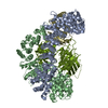
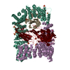
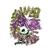
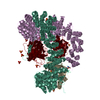
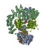
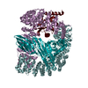
| Entry | Database: PDB / ID: 6owo | ||||||
|---|---|---|---|---|---|---|---|
| Title | CRYO-EM STRUCTURE OF PHOSPHORYLATED AP-2 CORE BOUND TO NECAP | ||||||
 Components Components |
| ||||||
 Keywords Keywords | ENDOCYTOSIS / AP2 / NECAP2 PROTEIN / CLATHRIN VESICLE / LIPID-BINDING / ADAPTOR / MEMBRANE / TRANSPORT / PHOSPHORYLATION | ||||||
| Function / homology |  Function and homology information Function and homology informationFormation of annular gap junctions / Gap junction degradation / clathrin vesicle coat / LDL clearance / WNT5A-dependent internalization of FZD2, FZD5 and ROR2 / WNT5A-dependent internalization of FZD2, FZD5 and ROR2 / WNT5A-dependent internalization of FZD4 / LDL clearance / Retrograde neurotrophin signalling / VLDLR internalisation and degradation ...Formation of annular gap junctions / Gap junction degradation / clathrin vesicle coat / LDL clearance / WNT5A-dependent internalization of FZD2, FZD5 and ROR2 / WNT5A-dependent internalization of FZD2, FZD5 and ROR2 / WNT5A-dependent internalization of FZD4 / LDL clearance / Retrograde neurotrophin signalling / VLDLR internalisation and degradation / Retrograde neurotrophin signalling / WNT5A-dependent internalization of FZD4 / clathrin coat / cardiac septum development / clathrin adaptor complex / extrinsic component of presynaptic endocytic zone membrane / MHC class II antigen presentation / Trafficking of GluR2-containing AMPA receptors / VLDLR internalisation and degradation / Recycling pathway of L1 / regulation of vesicle size / postsynaptic endocytic zone / postsynaptic neurotransmitter receptor internalization / AP-2 adaptor complex / Recycling pathway of L1 / Cargo recognition for clathrin-mediated endocytosis / membrane coat / Cargo recognition for clathrin-mediated endocytosis / clathrin coat assembly / Clathrin-mediated endocytosis / positive regulation of synaptic vesicle endocytosis / Clathrin-mediated endocytosis / clathrin-cargo adaptor activity / vesicle budding from membrane / clathrin-dependent endocytosis / MHC class II antigen presentation / signal sequence receptor activity / coronary vasculature development / positive regulation of protein localization to membrane / neurotransmitter secretion / regulation of hematopoietic stem cell differentiation / ventricular septum development / aorta development / low-density lipoprotein particle receptor binding / clathrin binding / Trafficking of GluR2-containing AMPA receptors / positive regulation of receptor internalization / positive regulation of endocytosis / negative regulation of protein localization to plasma membrane / synaptic vesicle endocytosis / vesicle-mediated transport / Neutrophil degranulation / clathrin-coated pit / phosphatidylinositol binding / secretory granule / protein serine/threonine kinase binding / kidney development / intracellular protein transport / receptor internalization / kinase binding / cytoplasmic side of plasma membrane / endocytosis / disordered domain specific binding / synaptic vesicle / protein transport / heart development / protein-containing complex assembly / cytoplasmic vesicle / transmembrane transporter binding / postsynapse / protein domain specific binding / lipid binding / synapse / protein kinase binding / protein-containing complex binding / glutamatergic synapse / mitochondrion / plasma membrane Similarity search - Function | ||||||
| Biological species |  | ||||||
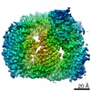
| Method | ELECTRON MICROSCOPY / single particle reconstruction / cryo EM / Resolution: 3.2 Å | ||||||
 Authors Authors | Partlow, E.A. / Baker, R.W. / Beacham, G.M. / Chappie, J.S. / Leschziner, A.E. / Hollopeter, G. | ||||||
| Funding support |  United States, 1items United States, 1items
| ||||||
 Citation Citation | Journal: Elife / Year: 2019 Title: A structural mechanism for phosphorylation-dependent inactivation of the AP2 complex. Authors: Edward A Partlow / Richard W Baker / Gwendolyn M Beacham / Joshua S Chappie / Andres E Leschziner / Gunther Hollopeter / Abstract: Endocytosis of transmembrane proteins is orchestrated by the AP2 clathrin adaptor complex. AP2 dwells in a closed, inactive state in the cytosol, but adopts an open, active conformation on the plasma ...Endocytosis of transmembrane proteins is orchestrated by the AP2 clathrin adaptor complex. AP2 dwells in a closed, inactive state in the cytosol, but adopts an open, active conformation on the plasma membrane. Membrane-activated complexes are also phosphorylated, but the significance of this mark is debated. We recently proposed that NECAP negatively regulates AP2 by binding open and phosphorylated complexes (Beacham et al., 2018). Here, we report high-resolution cryo-EM structures of NECAP bound to phosphorylated AP2. The site of AP2 phosphorylation is directly coordinated by residues of the NECAP PHear domain that are predicted from genetic screens in . Using membrane mimetics to generate conformationally open AP2, we find that a second domain of NECAP binds these complexes and cryo-EM reveals both domains of NECAP engaging closed, inactive AP2. Assays in vitro and in vivo confirm these domains cooperate to inactivate AP2. We propose that phosphorylation marks adaptors for inactivation. | ||||||
| History |
|
- Structure visualization
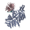
| Movie |
Movie viewer |
|---|---|
| Structure viewer | Molecule: MolmilJmol/JSmol |
- Downloads & links
-Download
| PDBx/mmCIF format | 6owo.cif.gz | 363.9 KB | Display | PDBx/mmCIF format |
|---|---|---|---|---|
| PDB format | pdb6owo.ent.gz | 289.2 KB | Display | PDB format |
| PDBx/mmJSON format | 6owo.json.gz | Tree view | PDBx/mmJSON format | |
| Others |  Other downloads Other downloads |
-Validation report
| Arichive directory | https://data.pdbj.org/pub/pdb/validation_reports/ow/6owoftp://data.pdbj.org/pub/pdb/validation_reports/ow/6owo | HTTPS FTP |
|---|
-Related structure data
| Related structure data |  20215MC 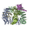 6oxlC M: map data used to model this data C: citing same article ( |
|---|---|
| Similar structure data |


-Links
 PDBj
PDBj


- Assembly
| Deposited unit | 
|
|---|---|
| 1 |
|
-Components
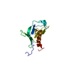
| #1: Protein | Mass: 69656.297 Da / Num. of mol.: 1 Source method: isolated from a genetically manipulated source Source: (gene. exp.)  |
|---|---|
| #2: Protein | Mass: 66953.195 Da / Num. of mol.: 1 Source method: isolated from a genetically manipulated source Source: (gene. exp.) |
| #3: Protein | Mass: 49806.621 Da / Num. of mol.: 1 Source method: isolated from a genetically manipulated source Source: (gene. exp.) |
| #4: Protein | Mass: 17038.688 Da / Num. of mol.: 1 Source method: isolated from a genetically manipulated source Source: (gene. exp.) |
| #5: Protein | Mass: 28629.941 Da / Num. of mol.: 1 Source method: isolated from a genetically manipulated source Source: (gene. exp.) |
| Has protein modification | Y |
-Experimental details
-Experiment
| Experiment | Method: ELECTRON MICROSCOPY |
|---|---|
| EM experiment | Aggregation state: PARTICLE / 3D reconstruction method: single particle reconstruction |
- Sample preparation
| Component | Name: PHOSPHORYLATED AP2-NECAP CO-COMPLEX / Type: COMPLEX / Entity ID: all / Source: MULTIPLE SOURCES |
|---|---|
| Molecular weight | Experimental value: NO |
| Source (natural) | Organism: |
| Source (recombinant) | Organism: |
| Buffer solution | pH: 8 |
| Specimen | Conc.: 1 mg/ml / Embedding applied: NO / Shadowing applied: NO / Staining applied: NO / Vitrification applied: YES |
| Specimen support | Details: GLOW DISCHARGE / Grid material: GOLD / Grid mesh size: 300 divisions/in. / Grid type: UltrAuFoil |
| Vitrification | Instrument: FEI VITROBOT MARK IV / Cryogen name: ETHANE / Humidity: 100 % / Chamber temperature: 277.15 K Details: 4 UL OF SAMPLE/GRID WAS BLOTTED FOR 4 SECONDS AND PLUNGE FROZEN IN LIQUID-NITROGEN COOLED ETHANE |
- Electron microscopy imaging
| Experimental equipment |  Model: Talos Arctica / Image courtesy: FEI Company |
|---|---|
| Microscopy | Model: FEI TALOS ARCTICA |
| Electron gun | Electron source:  FIELD EMISSION GUN / Accelerating voltage: 200 kV / Illumination mode: FLOOD BEAM FIELD EMISSION GUN / Accelerating voltage: 200 kV / Illumination mode: FLOOD BEAM |
| Electron lens | Mode: BRIGHT FIELD / Nominal magnification: 36000 X / Nominal defocus max: 2500 nm / Nominal defocus min: 600 nm / Cs: 2.7 mm |
| Specimen holder | Cryogen: NITROGEN |
| Image recording | Electron dose: 1 e/Å2 / Detector mode: SUPER-RESOLUTION / Film or detector model: GATAN K2 SUMMIT (4k x 4k) / Num. of real images: 944 |
- Processing
| EM software |
| ||||||||||||||||||||||||||||||||||||||||||||||||
|---|---|---|---|---|---|---|---|---|---|---|---|---|---|---|---|---|---|---|---|---|---|---|---|---|---|---|---|---|---|---|---|---|---|---|---|---|---|---|---|---|---|---|---|---|---|---|---|---|---|
| Image processing |
| ||||||||||||||||||||||||||||||||||||||||||||||||
| CTF correction |
| ||||||||||||||||||||||||||||||||||||||||||||||||
| Symmetry |
| ||||||||||||||||||||||||||||||||||||||||||||||||
| 3D reconstruction |
| ||||||||||||||||||||||||||||||||||||||||||||||||
| Atomic model building | B value: 63 / Protocol: RIGID BODY FIT / Space: REAL / Target criteria: MAXIMUM LIKELIHOOD Details: STARTING MODEL WAS GENERATED BY DOCKING PDB ENTRIES 2VGL AND 1TQZ INTO CRYO-EM DENSITY AND MANUALLY EDITING SEQUENCE AND STRUCTURAl CHANGES. MODEL REFINEMENT WAS PERFORMED USING ROSETTA AND ...Details: STARTING MODEL WAS GENERATED BY DOCKING PDB ENTRIES 2VGL AND 1TQZ INTO CRYO-EM DENSITY AND MANUALLY EDITING SEQUENCE AND STRUCTURAl CHANGES. MODEL REFINEMENT WAS PERFORMED USING ROSETTA AND PHENIX.REAL_SPACE_REFINE | ||||||||||||||||||||||||||||||||||||||||||||||||
| Atomic model building |
| ||||||||||||||||||||||||||||||||||||||||||||||||
| Refinement | Highest resolution: 3.2 Å |