National Institutes of Health/National Institute of General Medical Sciences (NIH/NIGMS)
R01 GM127548-01A1
United States
Citation
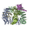
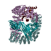
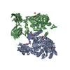
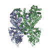
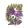
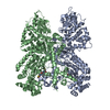
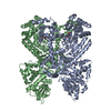
Journal: Elife / Year: 2019 Title: A structural mechanism for phosphorylation-dependent inactivation of the AP2 complex. Authors: Edward A Partlow / Richard W Baker / Gwendolyn M Beacham / Joshua S Chappie / Andres E Leschziner / Gunther Hollopeter / Abstract: Endocytosis of transmembrane proteins is orchestrated by the AP2 clathrin adaptor complex. AP2 dwells in a closed, inactive state in the cytosol, but adopts an open, active conformation on the plasma ...Endocytosis of transmembrane proteins is orchestrated by the AP2 clathrin adaptor complex. AP2 dwells in a closed, inactive state in the cytosol, but adopts an open, active conformation on the plasma membrane. Membrane-activated complexes are also phosphorylated, but the significance of this mark is debated. We recently proposed that NECAP negatively regulates AP2 by binding open and phosphorylated complexes (Beacham et al., 2018). Here, we report high-resolution cryo-EM structures of NECAP bound to phosphorylated AP2. The site of AP2 phosphorylation is directly coordinated by residues of the NECAP PHear domain that are predicted from genetic screens in . Using membrane mimetics to generate conformationally open AP2, we find that a second domain of NECAP binds these complexes and cryo-EM reveals both domains of NECAP engaging closed, inactive AP2. Assays in vitro and in vivo confirm these domains cooperate to inactivate AP2. We propose that phosphorylation marks adaptors for inactivation.
History
Deposition
May 10, 2019
-
Header (metadata) release
Aug 28, 2019
-
Map release
Sep 11, 2019
-
Update
Oct 16, 2024
-
Current status
Oct 16, 2024
Processing site: RCSB / Status: Released
-
Structure visualization
Movie
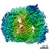
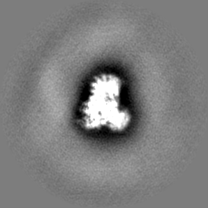
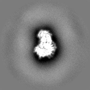
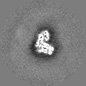
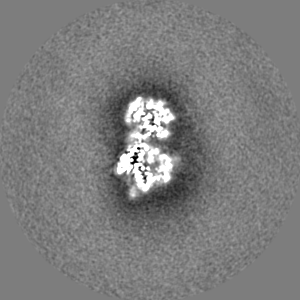
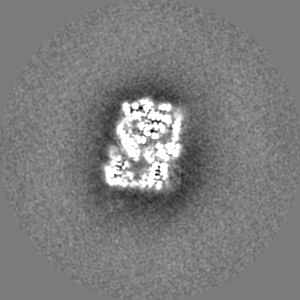
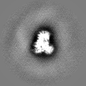
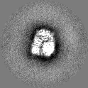
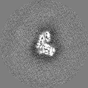
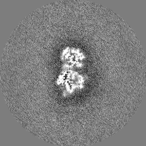
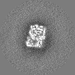
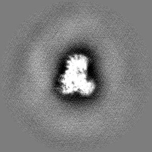
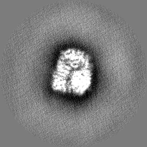
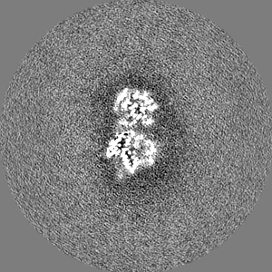
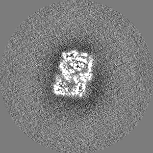
Surface view with section colored by density value
Cryogen name: ETHANE / Chamber humidity: 100 % / Chamber temperature: 277.15 K / Instrument: FEI VITROBOT MARK IV Details: 4 UL OF SAMPLE/GRID WAS BLOTTED FOR 4 SECONDS AND PLUNGE FROZEN IN LIQUID-NITROGEN COOLED ETHANE.
-
Electron microscopy
Microscope
FEI TALOS ARCTICA
Software
Name: Leginon
Image recording
Film or detector model: GATAN K2 SUMMIT (4k x 4k) / Detector mode: SUPER-RESOLUTION / Number real images: 944 / Average electron dose: 1.0 e/Å2
Electron beam
Acceleration voltage: 200 kV / Electron source: FIELD EMISSION GUN
source_name: PDB, initial_model_type: experimental model
Details
STARTING MODEL WAS GENERATED BY DOCKING PDB ENTRIES 2VGL AND 1TQZ INTO CRYO-EM DENSITY AND MANUALLY EDITING SEQUENCE AND STRUCTURAl CHANGES. MODEL REFINEMENT WAS PERFORMED USING ROSETTA AND PHENIX.REAL_SPACE_REFINE
Refinement
Space: REAL / Protocol: RIGID BODY FIT / Overall B value: 63 / Target criteria: MAXIMUM LIKELIHOOD
Output model
PDB-6owo: CRYO-EM STRUCTURE OF PHOSPHORYLATED AP-2 CORE BOUND TO NECAP
+
About Yorodumi
-
News
-
Feb 9, 2022. New format data for meta-information of EMDB entries
New format data for meta-information of EMDB entries
Version 3 of the EMDB header file is now the official format.
The previous official version 1.9 will be removed from the archive.
In the structure databanks used in Yorodumi, some data are registered as the other names, "COVID-19 virus" and "2019-nCoV". Here are the details of the virus and the list of structure data.
Jan 31, 2019. EMDB accession codes are about to change! (news from PDBe EMDB page)
EMDB accession codes are about to change! (news from PDBe EMDB page)
The allocation of 4 digits for EMDB accession codes will soon come to an end. Whilst these codes will remain in use, new EMDB accession codes will include an additional digit and will expand incrementally as the available range of codes is exhausted. The current 4-digit format prefixed with “EMD-” (i.e. EMD-XXXX) will advance to a 5-digit format (i.e. EMD-XXXXX), and so on. It is currently estimated that the 4-digit codes will be depleted around Spring 2019, at which point the 5-digit format will come into force.
The EM Navigator/Yorodumi systems omit the EMD- prefix.
Related info.:Q: What is EMD? / ID/Accession-code notation in Yorodumi/EM Navigator
Yorodumi is a browser for structure data from EMDB, PDB, SASBDB, etc.
This page is also the successor to EM Navigator detail page, and also detail information page/front-end page for Omokage search.
The word "yorodu" (or yorozu) is an old Japanese word meaning "ten thousand". "mi" (miru) is to see.
Related info.:EMDB / PDB / SASBDB / Comparison of 3 databanks / Yorodumi Search / Aug 31, 2016. New EM Navigator & Yorodumi / Yorodumi Papers / Jmol/JSmol / Function and homology information / Changes in new EM Navigator and Yorodumi
 Movie
Movie Controller


 Open data
 Basic information
 Map data
 Sample
 Keywords
 Function and homology information
 Authors
 United States, 1 items
  Citation
 Structure visualization
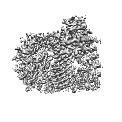
 Downloads & links
 emd_20215.png
 http://ftp.pdbj.org/pub/emdb/structures/EMD-20215
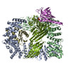


 Z (Sec.)
 Y (Row.)
 X (Col.)


 Sample components
 Processing
 Electron microscopy
 FIELD EMISSION GUN