 Movie
Movie Controller

[English] 日本語
 Yorodumi
- PDB-6m4w: Crystal structure of MBP fused split FKBP-FRB T2098L mutant in co... -
+ Open data

- Basic information
| Entry | Database: PDB / ID: 6m4w | ||||||
|---|---|---|---|---|---|---|---|
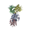
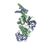
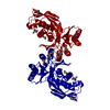
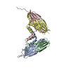
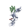
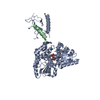
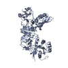
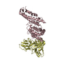
| Title | Crystal structure of MBP fused split FKBP-FRB T2098L mutant in complex with rapamycin | ||||||
 Components Components |
| ||||||
 Keywords Keywords | ISOMERASE / Rapamycin / complex / kinase | ||||||
| Function / homology |  Function and homology information Function and homology informationpositive regulation of SCF-dependent proteasomal ubiquitin-dependent catabolic process / RNA polymerase III type 2 promoter sequence-specific DNA binding / RNA polymerase III type 1 promoter sequence-specific DNA binding / positive regulation of cytoplasmic translational initiation / regulation of locomotor rhythm / T-helper 1 cell lineage commitment / positive regulation of pentose-phosphate shunt / positive regulation of wound healing, spreading of epidermal cells / macrolide binding / regulation of membrane permeability ...positive regulation of SCF-dependent proteasomal ubiquitin-dependent catabolic process / RNA polymerase III type 2 promoter sequence-specific DNA binding / RNA polymerase III type 1 promoter sequence-specific DNA binding / positive regulation of cytoplasmic translational initiation / regulation of locomotor rhythm / T-helper 1 cell lineage commitment / positive regulation of pentose-phosphate shunt / positive regulation of wound healing, spreading of epidermal cells / macrolide binding / regulation of membrane permeability / TORC2 complex / cellular response to leucine starvation / TFIIIC-class transcription factor complex binding / heart valve morphogenesis / voluntary musculoskeletal movement / activin receptor binding / negative regulation of lysosome organization / TORC1 complex / calcineurin-NFAT signaling cascade / regulation of skeletal muscle contraction by regulation of release of sequestered calcium ion / RNA polymerase III type 3 promoter sequence-specific DNA binding / positive regulation of transcription of nucleolar large rRNA by RNA polymerase I / positive regulation of keratinocyte migration / cytoplasmic side of membrane / regulation of osteoclast differentiation / energy reserve metabolic process / transforming growth factor beta receptor binding / TGFBR1 LBD Mutants in Cancer / MTOR signalling / regulation of lysosome organization / cellular response to L-leucine / Energy dependent regulation of mTOR by LKB1-AMPK / cellular response to nutrient / regulation of autophagosome assembly / type I transforming growth factor beta receptor binding / negative regulation of activin receptor signaling pathway / Amino acids regulate mTORC1 / signaling receptor inhibitor activity / heart trabecula formation / cellular response to methionine / negative regulation of cell size / TORC2 signaling / I-SMAD binding / cellular response to osmotic stress / regulation of amyloid precursor protein catabolic process / terminal cisterna / ryanodine receptor complex / cell projection organization / anoikis / inositol hexakisphosphate binding / cardiac muscle cell development / negative regulation of calcineurin-NFAT signaling cascade / negative regulation of protein localization to nucleus / positive regulation of ubiquitin-dependent protein catabolic process / regulation of myelination / positive regulation of transcription by RNA polymerase III / 'de novo' protein folding / negative regulation of macroautophagy / positive regulation of ruffle assembly / regulation of cell size / ventricular cardiac muscle tissue morphogenesis / FK506 binding / positive regulation of actin filament polymerization / positive regulation of myotube differentiation / Macroautophagy / Constitutive Signaling by AKT1 E17K in Cancer / oligodendrocyte differentiation / detection of maltose stimulus / maltose transport complex / germ cell development / positive regulation of oligodendrocyte differentiation / TORC1 signaling / TGF-beta receptor signaling activates SMADs / carbohydrate transport / TOR signaling / response to amino acid / behavioral response to pain / mTORC1-mediated signalling / Calcineurin activates NFAT / CD28 dependent PI3K/Akt signaling / HSF1-dependent transactivation / regulation of macroautophagy / regulation of immune response / positive regulation of translational initiation / carbohydrate transmembrane transporter activity / maltose binding / maltose transport / maltodextrin transmembrane transport / positive regulation of epithelial to mesenchymal transition / positive regulation of lipid biosynthetic process / heart morphogenesis / vascular endothelial cell response to laminar fluid shear stress / 'de novo' pyrimidine nucleobase biosynthetic process / regulation of cellular response to heat / positive regulation of lamellipodium assembly / ATP-binding cassette (ABC) transporter complex, substrate-binding subunit-containing / neuronal action potential / T cell costimulation / phagocytic vesicle / cardiac muscle contraction Similarity search - Function | ||||||
| Biological species |   Homo sapiens (human) Homo sapiens (human) | ||||||
| Method |  X-RAY DIFFRACTION / SYNCHROTRON / MOLECULAR REPLACEMENT / Resolution: 3.11 Å X-RAY DIFFRACTION / SYNCHROTRON / MOLECULAR REPLACEMENT / Resolution: 3.11 Å | ||||||
 Authors Authors | Kikuchi, M. / Wu, D. / Inoue, T. / Umehara, T. | ||||||
 Citation Citation | Journal: Nat.Methods / Year: 2020 Title: Rational design and implementation of a chemically inducible heterotrimerization system. Authors: Wu, H.D. / Kikuchi, M. / Dagliyan, O. / Aragaki, A.K. / Nakamura, H. / Dokholyan, N.V. / Umehara, T. / Inoue, T. | ||||||
| History |
|
- Structure visualization
| Structure viewer | Molecule: MolmilJmol/JSmol |
|---|
- Downloads & links
-Download
| PDBx/mmCIF format | 6m4w.cif.gz | 346.3 KB | Display | PDBx/mmCIF format |
|---|---|---|---|---|
| PDB format | pdb6m4w.ent.gz | 280.3 KB | Display | PDB format |
| PDBx/mmJSON format | 6m4w.json.gz | Tree view | PDBx/mmJSON format | |
| Others |  Other downloads Other downloads |
-Validation report
| Arichive directory | https://data.pdbj.org/pub/pdb/validation_reports/m4/6m4wftp://data.pdbj.org/pub/pdb/validation_reports/m4/6m4w | HTTPS FTP |
|---|
-Related structure data
| Related structure data |  6m4uC 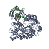 6m4vC  1fapS S: Starting model for refinement C: citing same article ( |
|---|---|
| Similar structure data |


-Links
 PDBj
PDBj


- Assembly
| Deposited unit | 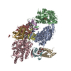
| ||||||||
|---|---|---|---|---|---|---|---|---|---|
| 1 | 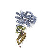
| ||||||||
| 2 | 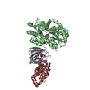
| ||||||||
| 3 | 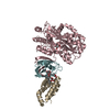
| ||||||||
| Unit cell |
|
-Components
-Protein , 3 types, 9 molecules ABCDEFGHI
| #1: Protein | Mass: 44074.879 Da / Num. of mol.: 3 / Mutation: D-288A, K-287A, E-198A, N-197A, K-131A Source method: isolated from a genetically manipulated source Details: This entity is chimeric. Organism is Homo sapiens and Eschrichia coli. Source: (gene. exp.) Homo sapiens (human)Strain: K-12 / Gene: malE, b4034, JW3994, FKBP1A, FKBP1, FKBP12 / Production host: References: UniProt: P0AEX9, UniProt: P62942, peptidylprolyl isomerase #2: Protein | Mass: 8486.715 Da / Num. of mol.: 3 Source method: isolated from a genetically manipulated source Source: (gene. exp.) Homo sapiens (human) / Gene: FKBP1A, FKBP1, FKBP12 / Production host: #3: Protein | Mass: 11447.091 Da / Num. of mol.: 3 / Mutation: T2098L Source method: isolated from a genetically manipulated source Source: (gene. exp.) Homo sapiens (human) / Gene: MTOR / Production host: References: UniProt: P42345, non-specific serine/threonine protein kinase |
|---|
-Sugars , 1 types, 3 molecules
| #4: Polysaccharide |   Source method: isolated from a genetically manipulated source Details: oligosaccharide / References: alpha-maltose |
|---|
-Non-polymers , 3 types, 195 molecules 


| #5: Chemical |  Mass: 914.172 Da / Num. of mol.: 3 / Source method: obtained synthetically / Formula: C51H79NO13 / Feature type: SUBJECT OF INVESTIGATION / Comment: antibiotic*YM Mass: 914.172 Da / Num. of mol.: 3 / Source method: obtained synthetically / Formula: C51H79NO13 / Feature type: SUBJECT OF INVESTIGATION / Comment: antibiotic*YM#6: Chemical | ChemComp-GOL / |  Mass: 92.094 Da / Num. of mol.: 1 / Source method: obtained synthetically / Formula: C3H8O3 Mass: 92.094 Da / Num. of mol.: 1 / Source method: obtained synthetically / Formula: C3H8O3#7: Water | ChemComp-HOH / | Mass: 18.015 Da / Num. of mol.: 191 / Source method: isolated from a natural source / Formula: H2O |
|---|
-Details
| Has ligand of interest | Y |
|---|
-Experimental details
-Experiment
| Experiment | Method: X-RAY DIFFRACTION / Number of used crystals: 1 |
|---|
- Sample preparation
| Crystal | Density Matthews: 2.95 Å3/Da / Density % sol: 58.3 % |
|---|---|
| Crystal grow | Temperature: 293 K / Method: vapor diffusion, sitting drop Details: 100 mM Tris-HCl buffer (pH 7.0), 200 mM calcium acetate and 20% (w/v) PEG 3000 |
-Data collection
| Diffraction | Mean temperature: 100 K / Serial crystal experiment: N |
|---|---|
| Diffraction source | Source: SYNCHROTRON / Site: SPring-8  / Beamline: BL26B2 / Wavelength: 1 Å / Beamline: BL26B2 / Wavelength: 1 Å |
| Detector | Type: RAYONIX MX225-HS / Detector: CCD / Date: Dec 10, 2019 |
| Radiation | Protocol: SINGLE WAVELENGTH / Monochromatic (M) / Laue (L): M / Scattering type: x-ray |
| Radiation wavelength | Wavelength: 1 Å / Relative weight: 1 |
| Reflection | Resolution: 3.11→48.6 Å / Num. obs: 42250 / % possible obs: 100 % / Redundancy: 14.8 % / CC1/2: 0.992 / Net I/σ(I): 9.5 |
| Reflection shell | Resolution: 3.11→3.23 Å / Num. unique obs: 4337 / CC1/2: 0.522 |
- Processing
| Software |
| ||||||||||||||||||||||||||||||||||||||||||||||||||||||||||||||||||||||||||||||||||||||||||||||||||||||||||||||||||||||||||||||||||||||||||||||||||||||||||||||||||||||||||||||||||||||
|---|---|---|---|---|---|---|---|---|---|---|---|---|---|---|---|---|---|---|---|---|---|---|---|---|---|---|---|---|---|---|---|---|---|---|---|---|---|---|---|---|---|---|---|---|---|---|---|---|---|---|---|---|---|---|---|---|---|---|---|---|---|---|---|---|---|---|---|---|---|---|---|---|---|---|---|---|---|---|---|---|---|---|---|---|---|---|---|---|---|---|---|---|---|---|---|---|---|---|---|---|---|---|---|---|---|---|---|---|---|---|---|---|---|---|---|---|---|---|---|---|---|---|---|---|---|---|---|---|---|---|---|---|---|---|---|---|---|---|---|---|---|---|---|---|---|---|---|---|---|---|---|---|---|---|---|---|---|---|---|---|---|---|---|---|---|---|---|---|---|---|---|---|---|---|---|---|---|---|---|---|---|---|---|
| Refinement | Method to determine structure: MOLECULAR REPLACEMENT Starting model: 1FAP Resolution: 3.11→48.65 Å / Cor.coef. Fo:Fc: 0.915 / Cor.coef. Fo:Fc free: 0.88 / SU B: 27.493 / SU ML: 0.45 / Cross valid method: THROUGHOUT / ESU R Free: 0.517 Details: HYDROGENS HAVE BEEN USED IF PRESENT IN THE INPUT SF FILE CONTAINS FRIEDEL PAIRS UNDER I/F_MINUS AND I/F_PLUS COLUMNS.
| ||||||||||||||||||||||||||||||||||||||||||||||||||||||||||||||||||||||||||||||||||||||||||||||||||||||||||||||||||||||||||||||||||||||||||||||||||||||||||||||||||||||||||||||||||||||
| Solvent computation | Ion probe radii: 0.8 Å / Shrinkage radii: 0.8 Å / VDW probe radii: 1.2 Å | ||||||||||||||||||||||||||||||||||||||||||||||||||||||||||||||||||||||||||||||||||||||||||||||||||||||||||||||||||||||||||||||||||||||||||||||||||||||||||||||||||||||||||||||||||||||
| Displacement parameters | Biso mean: 70.304 Å2
| ||||||||||||||||||||||||||||||||||||||||||||||||||||||||||||||||||||||||||||||||||||||||||||||||||||||||||||||||||||||||||||||||||||||||||||||||||||||||||||||||||||||||||||||||||||||
| Refinement step | Cycle: 1 / Resolution: 3.11→48.65 Å
| ||||||||||||||||||||||||||||||||||||||||||||||||||||||||||||||||||||||||||||||||||||||||||||||||||||||||||||||||||||||||||||||||||||||||||||||||||||||||||||||||||||||||||||||||||||||
| Refine LS restraints |
|