 Movie
Movie Controller

[English] 日本語
 Yorodumi
- PDB-6k67: Application of anti-helix antibodies in protein structure determi... -
+ Open data

- Basic information
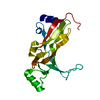
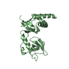
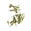
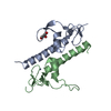
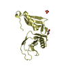
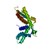
| Entry | Database: PDB / ID: 6k67 | |||||||||
|---|---|---|---|---|---|---|---|---|---|---|
| Title | Application of anti-helix antibodies in protein structure determination (9011-3LRH) | |||||||||
 Components Components |
| |||||||||
 Keywords Keywords | STRUCTURAL PROTEIN / antibody / protein design | |||||||||
| Function / homology |  Function and homology information Function and homology informationnegative regulation of high voltage-gated calcium channel activity / positive regulation of cyclic-nucleotide phosphodiesterase activity / negative regulation of calcium ion export across plasma membrane / regulation of cardiac muscle cell action potential / positive regulation of ryanodine-sensitive calcium-release channel activity / regulation of cell communication by electrical coupling involved in cardiac conduction / negative regulation of peptidyl-threonine phosphorylation / protein phosphatase activator activity / positive regulation of phosphoprotein phosphatase activity / adenylate cyclase binding ...negative regulation of high voltage-gated calcium channel activity / positive regulation of cyclic-nucleotide phosphodiesterase activity / negative regulation of calcium ion export across plasma membrane / regulation of cardiac muscle cell action potential / positive regulation of ryanodine-sensitive calcium-release channel activity / regulation of cell communication by electrical coupling involved in cardiac conduction / negative regulation of peptidyl-threonine phosphorylation / protein phosphatase activator activity / positive regulation of phosphoprotein phosphatase activity / adenylate cyclase binding / catalytic complex / detection of calcium ion / regulation of cardiac muscle contraction / negative regulation of ryanodine-sensitive calcium-release channel activity / regulation of cardiac muscle contraction by regulation of the release of sequestered calcium ion / regulation of release of sequestered calcium ion into cytosol by sarcoplasmic reticulum / positive regulation of protein dephosphorylation / regulation of calcium-mediated signaling / titin binding / positive regulation of protein autophosphorylation / voltage-gated potassium channel complex / sperm midpiece / calcium channel complex / substantia nigra development / adenylate cyclase activator activity / regulation of heart rate / sarcomere / protein serine/threonine kinase activator activity / regulation of cytokinesis / positive regulation of peptidyl-threonine phosphorylation / positive regulation of protein serine/threonine kinase activity / spindle microtubule / spindle pole / response to calcium ion / G2/M transition of mitotic cell cycle / calcium-dependent protein binding / myelin sheath / vesicle / transmembrane transporter binding / G protein-coupled receptor signaling pathway / centrosome / calcium ion binding / protein kinase binding / protein-containing complex / nucleus / plasma membrane / cytoplasm Similarity search - Function | |||||||||
| Biological species |  Homo sapiens (human) Homo sapiens (human) | |||||||||
| Method |  X-RAY DIFFRACTION / SYNCHROTRON / MOLECULAR REPLACEMENT / Resolution: 1.95 Å X-RAY DIFFRACTION / SYNCHROTRON / MOLECULAR REPLACEMENT / Resolution: 1.95 Å | |||||||||
 Authors Authors | Lee, J.O. / Jin, M.S. / Kim, J.W. / Kim, S. / Lee, H. / Cho, G.Y. | |||||||||
| Funding support |  Korea, Republic Of, 2items Korea, Republic Of, 2items
| |||||||||
 Citation Citation | Journal: Proc.Natl.Acad.Sci.USA / Year: 2019 Title: Application of antihelix antibodies in protein structure determination. Authors: Kim, J.W. / Kim, S. / Lee, H. / Cho, G. / Kim, S.C. / Lee, H. / Jin, M.S. / Lee, J.O. | |||||||||
| History |
|
- Structure visualization
| Structure viewer | Molecule: MolmilJmol/JSmol |
|---|
- Downloads & links
-Download
| PDBx/mmCIF format | 6k67.cif.gz | 94.5 KB | Display | PDBx/mmCIF format |
|---|---|---|---|---|
| PDB format | pdb6k67.ent.gz | 69.6 KB | Display | PDB format |
| PDBx/mmJSON format | 6k67.json.gz | Tree view | PDBx/mmJSON format | |
| Others |  Other downloads Other downloads |
-Validation report
| Summary document | 6k67_validation.pdf.gz | 2.2 MB | Display | wwPDB validaton report |
|---|---|---|---|---|
| Full document | 6k67_full_validation.pdf.gz | 2.2 MB | Display | |
| Data in XML | 6k67_validation.xml.gz | 18.2 KB | Display | |
| Data in CIF | 6k67_validation.cif.gz | 26.1 KB | Display | |
| Arichive directory | https://data.pdbj.org/pub/pdb/validation_reports/k6/6k67ftp://data.pdbj.org/pub/pdb/validation_reports/k6/6k67 | HTTPS FTP |
-Related structure data
| Related structure data | 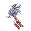 6k3mC 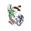 6k64C  6k65C 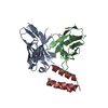 6k68C 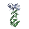 6k69C 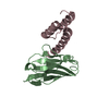 6k6aC 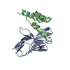 6k6bC  2w73S  3lrhS S: Starting model for refinement C: citing same article ( |
|---|---|
| Similar structure data |


-Links
 PDBj
PDBj

- Assembly
| Deposited unit | 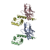
| ||||||||
|---|---|---|---|---|---|---|---|---|---|
| 1 | 
| ||||||||
| 2 | 
| ||||||||
| Unit cell |
|
-Components
| #1: Protein | Mass: 14203.453 Da / Num. of mol.: 2 Source method: isolated from a genetically manipulated source Source: (gene. exp.) Homo sapiens (human) / Production host:  #2: Protein | Mass: 11685.125 Da / Num. of mol.: 2 Source method: isolated from a genetically manipulated source Source: (gene. exp.) Homo sapiens (human) / Production host: #3: Chemical | ChemComp-CA /   Mass: 40.078 Da / Num. of mol.: 4 / Source method: obtained synthetically / Formula: Ca / Feature type: SUBJECT OF INVESTIGATION Mass: 40.078 Da / Num. of mol.: 4 / Source method: obtained synthetically / Formula: Ca / Feature type: SUBJECT OF INVESTIGATION#4: Water | ChemComp-HOH / |  Mass: 18.015 Da / Num. of mol.: 238 / Source method: isolated from a natural source / Formula: H2O Mass: 18.015 Da / Num. of mol.: 238 / Source method: isolated from a natural source / Formula: H2O |
|---|
-Experimental details
-Experiment
| Experiment | Method: X-RAY DIFFRACTION / Number of used crystals: 1 |
|---|
- Sample preparation
| Crystal | Density Matthews: 2.3 Å3/Da / Density % sol: 46.62 % |
|---|---|
| Crystal grow | Temperature: 296 K / Method: vapor diffusion, sitting drop / Details: 50% PEG 400, 0.2M CaCl2, 0.1M HEPES pH 7.5 |
-Data collection
| Diffraction | Mean temperature: 103 K / Serial crystal experiment: N |
|---|---|
| Diffraction source | Source: SYNCHROTRON / Site: PAL/PLS / Beamline: 7A (6B, 6C1) / Wavelength: 0.979 Å |
| Detector | Type: ADSC QUANTUM 315r / Detector: CCD / Date: Jul 19, 2016 |
| Radiation | Protocol: SINGLE WAVELENGTH / Monochromatic (M) / Laue (L): M / Scattering type: x-ray |
| Radiation wavelength | Wavelength: 0.979 Å / Relative weight: 1 |
| Reflection | Resolution: 1.9→50 Å / Num. obs: 33481 / % possible obs: 99.7 % / Redundancy: 5.1 % / Rpim(I) all: 0.043 / Net I/σ(I): 27.3 |
| Reflection shell | Resolution: 1.9→2 Å / Num. unique obs: 3373 / Rpim(I) all: 0.26 |
- Processing
| Software |
| ||||||||||||||||||||||||||||||||||||||||||||||||||||||||||||||||||||||||||||||||||||||||||||||||||||||||||||||||||||||||||||||||||||||||||||||||||||||||||||||||||||||||||||||||||||||
|---|---|---|---|---|---|---|---|---|---|---|---|---|---|---|---|---|---|---|---|---|---|---|---|---|---|---|---|---|---|---|---|---|---|---|---|---|---|---|---|---|---|---|---|---|---|---|---|---|---|---|---|---|---|---|---|---|---|---|---|---|---|---|---|---|---|---|---|---|---|---|---|---|---|---|---|---|---|---|---|---|---|---|---|---|---|---|---|---|---|---|---|---|---|---|---|---|---|---|---|---|---|---|---|---|---|---|---|---|---|---|---|---|---|---|---|---|---|---|---|---|---|---|---|---|---|---|---|---|---|---|---|---|---|---|---|---|---|---|---|---|---|---|---|---|---|---|---|---|---|---|---|---|---|---|---|---|---|---|---|---|---|---|---|---|---|---|---|---|---|---|---|---|---|---|---|---|---|---|---|---|---|---|---|
| Refinement | Method to determine structure: MOLECULAR REPLACEMENT Starting model: 3LRH, 2W73 Resolution: 1.95→27.38 Å / Cor.coef. Fo:Fc: 0.957 / Cor.coef. Fo:Fc free: 0.941 / SU B: 4.074 / SU ML: 0.111 / Cross valid method: THROUGHOUT / ESU R: 0.164 / ESU R Free: 0.148 / Details: HYDROGENS HAVE BEEN ADDED IN THE RIDING POSITIONS
| ||||||||||||||||||||||||||||||||||||||||||||||||||||||||||||||||||||||||||||||||||||||||||||||||||||||||||||||||||||||||||||||||||||||||||||||||||||||||||||||||||||||||||||||||||||||
| Solvent computation | Ion probe radii: 0.8 Å / Shrinkage radii: 0.8 Å / VDW probe radii: 1.2 Å | ||||||||||||||||||||||||||||||||||||||||||||||||||||||||||||||||||||||||||||||||||||||||||||||||||||||||||||||||||||||||||||||||||||||||||||||||||||||||||||||||||||||||||||||||||||||
| Displacement parameters | Biso mean: 36.547 Å2
| ||||||||||||||||||||||||||||||||||||||||||||||||||||||||||||||||||||||||||||||||||||||||||||||||||||||||||||||||||||||||||||||||||||||||||||||||||||||||||||||||||||||||||||||||||||||
| Refinement step | Cycle: 1 / Resolution: 1.95→27.38 Å
| ||||||||||||||||||||||||||||||||||||||||||||||||||||||||||||||||||||||||||||||||||||||||||||||||||||||||||||||||||||||||||||||||||||||||||||||||||||||||||||||||||||||||||||||||||||||
| Refine LS restraints |
|