 Movie
Movie Controller

[English] 日本語
 Yorodumi
- PDB-3ktm: Structure of the Heparin-induced E1-Dimer of the Amyloid Precurso... -
+ Open data

- Basic information
| Entry | Database: PDB / ID: 3ktm | ||||||
|---|---|---|---|---|---|---|---|
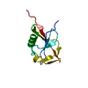
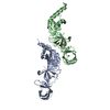
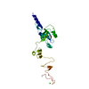
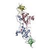
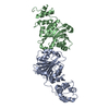
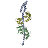
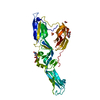
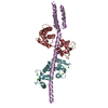
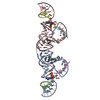
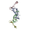
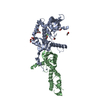
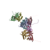
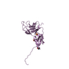
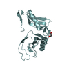
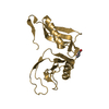
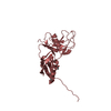
| Title | Structure of the Heparin-induced E1-Dimer of the Amyloid Precursor Protein (APP) | ||||||
 Components Components | Amyloid beta A4 protein | ||||||
 Keywords Keywords | CELL ADHESION / SIGNALING PROTEIN / Protein Structure / Alzheimer disease / Amyloid / Amyloidosis / Apoptosis / Copper / Disease mutation / Disulfide bond / Endocytosis / Heparin-binding / Metal-binding / Neurodegeneration / Notch signaling pathway / Proteoglycan / Zinc | ||||||
| Function / homology |  Function and homology information Function and homology informationamyloid-beta complex / growth cone lamellipodium / cellular response to norepinephrine stimulus / collateral sprouting in absence of injury / growth cone filopodium / microglia development / Formyl peptide receptors bind formyl peptides and many other ligands / regulation of Wnt signaling pathway / axo-dendritic transport / axon midline choice point recognition ...amyloid-beta complex / growth cone lamellipodium / cellular response to norepinephrine stimulus / collateral sprouting in absence of injury / growth cone filopodium / microglia development / Formyl peptide receptors bind formyl peptides and many other ligands / regulation of Wnt signaling pathway / axo-dendritic transport / axon midline choice point recognition / hippocampal neuron apoptotic process / regulation of synapse structure or activity / astrocyte activation involved in immune response / NMDA selective glutamate receptor signaling pathway / regulation of spontaneous synaptic transmission / mating behavior / growth factor receptor binding / peptidase activator activity / Insertion of tail-anchored proteins into the endoplasmic reticulum membrane / positive regulation of amyloid fibril formation / Golgi-associated vesicle / PTB domain binding / astrocyte projection / Lysosome Vesicle Biogenesis / Deregulated CDK5 triggers multiple neurodegenerative pathways in Alzheimer's disease models / neuron remodeling / nuclear envelope lumen / TRAF6 mediated NF-kB activation / dendrite development / positive regulation of protein metabolic process / signaling receptor activator activity / negative regulation of long-term synaptic potentiation / transition metal ion binding / Advanced glycosylation endproduct receptor signaling / The NLRP3 inflammasome / modulation of excitatory postsynaptic potential / regulation of multicellular organism growth / main axon / intracellular copper ion homeostasis / ECM proteoglycans / response to insulin-like growth factor stimulus / positive regulation of T cell migration / regulation of presynapse assembly / neuronal dense core vesicle / Purinergic signaling in leishmaniasis infection / cellular response to manganese ion / Notch signaling pathway / positive regulation of chemokine production / swimming behavior / neuron projection maintenance / extracellular matrix organization / clathrin-coated pit / positive regulation of mitotic cell cycle / axonogenesis / Mitochondrial protein degradation / ionotropic glutamate receptor signaling pathway / platelet alpha granule lumen / astrocyte activation / positive regulation of calcium-mediated signaling / response to interleukin-1 / regulation of neuron apoptotic process / cellular response to cAMP / cellular response to copper ion / positive regulation of glycolytic process / endosome lumen / trans-Golgi network membrane / positive regulation of interleukin-1 beta production / protein serine/threonine kinase binding / dendritic shaft / positive regulation of long-term synaptic potentiation / learning / central nervous system development / Post-translational protein phosphorylation / adult locomotory behavior / serine-type endopeptidase inhibitor activity / locomotory behavior / microglial cell activation / cellular response to nerve growth factor stimulus / positive regulation of non-canonical NF-kappaB signal transduction / TAK1-dependent IKK and NF-kappa-B activation / synapse organization / visual learning / recycling endosome / positive regulation of interleukin-6 production / positive regulation of JNK cascade / Golgi lumen / response to lead ion / regulation of long-term neuronal synaptic plasticity / cognition / Regulation of Insulin-like Growth Factor (IGF) transport and uptake by Insulin-like Growth Factor Binding Proteins (IGFBPs) / cellular response to amyloid-beta / endocytosis / neuron projection development / positive regulation of tumor necrosis factor production / positive regulation of inflammatory response / calcium ion transport / Platelet degranulation / regulation of translation / heparin binding / regulation of gene expression Similarity search - Function | ||||||
| Biological species |  Homo sapiens (human) Homo sapiens (human) | ||||||
| Method |  X-RAY DIFFRACTION / SYNCHROTRON / MOLECULAR REPLACEMENT / Resolution: 2.7 Å X-RAY DIFFRACTION / SYNCHROTRON / MOLECULAR REPLACEMENT / Resolution: 2.7 Å | ||||||
 Authors Authors | Dahms, S.O. / Hoefgen, S. / Roeser, D. / Schlott, B. / Guhrs, K.H. / Than, M.E. | ||||||
 Citation Citation | Journal: Proc.Natl.Acad.Sci.USA / Year: 2010 Title: Structure and biochemical analysis of the heparin-induced E1 dimer of the amyloid precursor protein. Authors: Dahms, S.O. / Hoefgen, S. / Roeser, D. / Schlott, B. / Guhrs, K.H. / Than, M.E. | ||||||
| History |
|
- Structure visualization
| Structure viewer | Molecule: MolmilJmol/JSmol |
|---|
- Downloads & links
-Download
| PDBx/mmCIF format | 3ktm.cif.gz | 292.5 KB | Display | PDBx/mmCIF format |
|---|---|---|---|---|
| PDB format | pdb3ktm.ent.gz | 237.1 KB | Display | PDB format |
| PDBx/mmJSON format | 3ktm.json.gz | Tree view | PDBx/mmJSON format | |
| Others |  Other downloads Other downloads |
-Validation report
| Arichive directory | https://data.pdbj.org/pub/pdb/validation_reports/kt/3ktmftp://data.pdbj.org/pub/pdb/validation_reports/kt/3ktm | HTTPS FTP |
|---|
-Related structure data

-Links
 PDBj
PDBj


- Assembly
-Components
| #1: Protein | Mass: 21762.080 Da / Num. of mol.: 8 / Fragment: UNP residues 18-190 Source method: isolated from a genetically manipulated source Source: (gene. exp.) Homo sapiens (human) / Gene: APP, A4, AD1 / Plasmid: pET22b / Production host:  #2: Chemical | ChemComp-BU4 / (   Mass: 90.121 Da / Num. of mol.: 8 / Source method: obtained synthetically / Formula: C4H10O2 Mass: 90.121 Da / Num. of mol.: 8 / Source method: obtained synthetically / Formula: C4H10O2#3: Chemical | ChemComp-SO4 /   Mass: 96.063 Da / Num. of mol.: 5 / Source method: obtained synthetically / Formula: SO4 Mass: 96.063 Da / Num. of mol.: 5 / Source method: obtained synthetically / Formula: SO4#4: Chemical | ChemComp-ACT /   Mass: 59.044 Da / Num. of mol.: 4 / Source method: obtained synthetically / Formula: C2H3O2 Mass: 59.044 Da / Num. of mol.: 4 / Source method: obtained synthetically / Formula: C2H3O2#5: Water | ChemComp-HOH / |  Mass: 18.015 Da / Num. of mol.: 282 / Source method: isolated from a natural source / Formula: H2O Mass: 18.015 Da / Num. of mol.: 282 / Source method: isolated from a natural source / Formula: H2OHas protein modification | Y | |
|---|
-Experimental details
-Experiment
| Experiment | Method: X-RAY DIFFRACTION / Number of used crystals: 1 |
|---|
- Sample preparation
| Crystal | Density Matthews: 4.02 Å3/Da / Density % sol: 69.44 % |
|---|---|
| Crystal grow | Temperature: 293 K / Method: vapor diffusion, sitting drop / pH: 5 Details: pH 5.0, VAPOR DIFFUSION, SITTING DROP, temperature 293K |
-Data collection
| Diffraction | Mean temperature: 100 K |
|---|---|
| Diffraction source | Source: SYNCHROTRON / Site: BESSY  / Beamline: 14.1 / Wavelength: 0.91841 Å / Beamline: 14.1 / Wavelength: 0.91841 Å |
| Detector | Type: MARMOSAIC 225 mm CCD / Detector: CCD / Date: Dec 19, 2008 Details: Double crystal monochromator with 2 sets of mirrors |
| Radiation | Protocol: SINGLE WAVELENGTH / Monochromatic (M) / Laue (L): M / Scattering type: x-ray |
| Radiation wavelength | Wavelength: 0.91841 Å / Relative weight: 1 |
| Reflection | Resolution: 2.7→29 Å / Num. obs: 74422 / % possible obs: 99.8 % / Redundancy: 2.1 % / Biso Wilson estimate: 69.152 Å2 / Rmerge(I) obs: 0.066 / Rsym value: 0.066 / Net I/σ(I): 10.8 |
| Reflection shell | Resolution: 2.7→2.85 Å / % possible obs: 99.7 % / Redundancy: 2 % / Rmerge(I) obs: 0.403 / Mean I/σ(I) obs: 2.1 / Num. unique all: 10822 |
- Processing
| Software |
| ||||||||||||||||||||
|---|---|---|---|---|---|---|---|---|---|---|---|---|---|---|---|---|---|---|---|---|---|
| Refinement | Method to determine structure: MOLECULAR REPLACEMENT Starting model: 1MWP, 2FJZ Resolution: 2.7→29 Å
| ||||||||||||||||||||
| Displacement parameters | Biso mean: 48.6 Å2 | ||||||||||||||||||||
| Refinement step | Cycle: LAST / Resolution: 2.7→29 Å
| ||||||||||||||||||||
| Refine LS restraints |
|
