Protocol: SINGLE WAVELENGTH / Monochromatic (M) / Laue (L): M / Scattering type: x-ray
Radiation wavelength
Wavelength: 0.98 Å / Relative weight: 1
Reflection
Resolution: 2.95→50 Å / Num. obs: 15977 / % possible obs: 99.57 % / Redundancy: 10.2 % / Rsym value: 0.152 / Net I/σ(I): 27.76
Reflection shell
Resolution: 2.95→3.026 Å / Redundancy: 10 % / Mean I/σ(I) obs: 4.25 / Rsym value: 0.596 / % possible all: 100
-
Processing
Software
Name
Version
Classification
REFMAC
5.8.0189
refinement
XDS
datareduction
SCALA
datascaling
Auto-Rickshaw
phasing
Refinement
Resolution: 2.95→38.4 Å / Cor.coef. Fo:Fc: 0.943 / Cor.coef. Fo:Fc free: 0.891 / SU B: 21.065 / SU ML: 0.389 / Cross valid method: THROUGHOUT / ESU R Free: 0.497 / Details: HYDROGENS HAVE BEEN ADDED IN THE RIDING POSITIONS
Rfactor
Num. reflection
% reflection
Selection details
Rfree
0.29418
775
6 %
RANDOM
Rwork
0.20059
-
-
-
obs
0.20622
12167
99.55 %
-
Solvent computation
Ion probe radii: 0.8 Å / Shrinkage radii: 0.8 Å / VDW probe radii: 1.2 Å
 Movie
Movie Controller

 Yorodumi
 Open data
 Basic information
 Components
 Keywords
 Function and homology information
 X-RAY DIFFRACTION /
  Authors
 India, 2items
  Citation
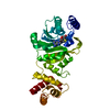
 Structure visualization
 Downloads & links
 Other downloads
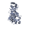


 PDBj
PDBj
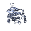
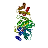
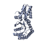
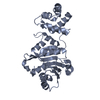
 Assembly
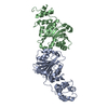

 Mass: 18.015 Da / Num. of mol.: 11 / Source method: isolated from a natural source / Formula: H2O
 Sample preparation
 / Beamline: ID29 / Wavelength: 0.98 Å
 Processing