 Movie
Movie Controller


+ Open data

- Basic information
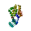
| Entry | Database: PDB / ID: 6k01 | ||||||
|---|---|---|---|---|---|---|---|
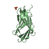
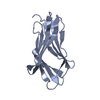
| Title | Crystal structure of xH2A-H2B | ||||||
 Components Components |
| ||||||
 Keywords Keywords | CHAPERONE / H2A / H2B | ||||||
| Function / homology |  Function and homology information Function and homology informationstructural constituent of chromatin / nucleosome / heterochromatin formation / protein heterodimerization activity / DNA binding / nucleus Similarity search - Function | ||||||
| Biological species | |||||||
| Method |  X-RAY DIFFRACTION / SYNCHROTRON / MOLECULAR REPLACEMENT / Resolution: 2.839 Å X-RAY DIFFRACTION / SYNCHROTRON / MOLECULAR REPLACEMENT / Resolution: 2.839 Å | ||||||
 Authors Authors | Liu, Y.R. | ||||||
 Citation Citation | Journal: Structure / Year: 2019 Title: Crystal structure of xlH2A-H2B Authors: Liu, Y.R. | ||||||
| History |
|
- Structure visualization
| Structure viewer | Molecule: MolmilJmol/JSmol |
|---|
- Downloads & links
-Download
| PDBx/mmCIF format | 6k01.cif.gz | 78.7 KB | Display | PDBx/mmCIF format |
|---|---|---|---|---|
| PDB format | pdb6k01.ent.gz | 57.5 KB | Display | PDB format |
| PDBx/mmJSON format | 6k01.json.gz | Tree view | PDBx/mmJSON format | |
| Others |  Other downloads Other downloads |
-Validation report
| Arichive directory | https://data.pdbj.org/pub/pdb/validation_reports/k0/6k01ftp://data.pdbj.org/pub/pdb/validation_reports/k0/6k01 | HTTPS FTP |
|---|
-Related structure data
| Related structure data | 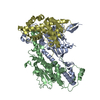 6k00C 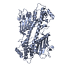 6k02C 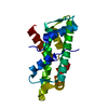 6k03C 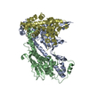 6k09C 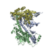 6k0cC 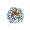 1aoiS S: Starting model for refinement C: citing same article ( |
|---|---|
| Similar structure data |


-Links
 PDBj
PDBj


- Assembly
| Deposited unit | 
| ||||||||
|---|---|---|---|---|---|---|---|---|---|
| 1 |
| ||||||||
| Unit cell |
|
-Components
| #1: Protein | Mass: 10133.729 Da / Num. of mol.: 1 Source method: isolated from a genetically manipulated source Source: (gene. exp.)  |
|---|---|
| #2: Protein | Mass: 11179.959 Da / Num. of mol.: 1 / Mutation: S30T Source method: isolated from a genetically manipulated source Source: (gene. exp.) |
-Experimental details
-Experiment
| Experiment | Method: X-RAY DIFFRACTION / Number of used crystals: 1 |
|---|
- Sample preparation
| Crystal | Density Matthews: 2 Å3/Da / Density % sol: 38.56 % |
|---|---|
| Crystal grow | Temperature: 291 K / Method: vapor diffusion, sitting drop Details: 0.2M Ammonium sulfate, 0.1M HEPES pH=7.5, 25% PEG 3350 |
-Data collection
| Diffraction | Mean temperature: 100 K / Serial crystal experiment: N |
|---|---|
| Diffraction source | Source: SYNCHROTRON / Site: SSRF  / Beamline: BL19U1 / Wavelength: 0.9793 Å / Beamline: BL19U1 / Wavelength: 0.9793 Å |
| Detector | Type: DECTRIS PILATUS3 6M / Detector: PIXEL / Date: Jun 4, 2018 |
| Radiation | Protocol: SINGLE WAVELENGTH / Monochromatic (M) / Laue (L): M / Scattering type: x-ray |
| Radiation wavelength | Wavelength: 0.9793 Å / Relative weight: 1 |
| Reflection | Resolution: 2.839→40.64 Å / Num. obs: 3756 / % possible obs: 99.6 % / Redundancy: 4.4 % / Net I/σ(I): 12.7 |
| Reflection shell | Resolution: 2.84→2.94 Å / Rmerge(I) obs: 0.831 / Num. unique obs: 3756 |
- Processing
| Software |
| |||||||||||||||||||||||||||||||||||||||||||||||||||||||||||||||||||||||||||
|---|---|---|---|---|---|---|---|---|---|---|---|---|---|---|---|---|---|---|---|---|---|---|---|---|---|---|---|---|---|---|---|---|---|---|---|---|---|---|---|---|---|---|---|---|---|---|---|---|---|---|---|---|---|---|---|---|---|---|---|---|---|---|---|---|---|---|---|---|---|---|---|---|---|---|---|---|
| Refinement | Method to determine structure: MOLECULAR REPLACEMENT Starting model: 1AOI Resolution: 2.839→40.637 Å / SU ML: 0.48 / Cross valid method: THROUGHOUT / σ(F): 2 / Phase error: 31.85 / Stereochemistry target values: ML Details: SF FILE CONTAINS FRIEDEL PAIRS UNDER I/F_MINUS AND I/F_PLUS COLUMNS.
| |||||||||||||||||||||||||||||||||||||||||||||||||||||||||||||||||||||||||||
| Solvent computation | Shrinkage radii: 0.9 Å / VDW probe radii: 1.11 Å / Solvent model: FLAT BULK SOLVENT MODEL | |||||||||||||||||||||||||||||||||||||||||||||||||||||||||||||||||||||||||||
| Displacement parameters | Biso max: 115.82 Å2 / Biso mean: 46.6349 Å2 / Biso min: 21 Å2 | |||||||||||||||||||||||||||||||||||||||||||||||||||||||||||||||||||||||||||
| Refinement step | Cycle: final / Resolution: 2.839→40.637 Å
| |||||||||||||||||||||||||||||||||||||||||||||||||||||||||||||||||||||||||||
| LS refinement shell | Refine-ID: X-RAY DIFFRACTION / Rfactor Rfree error: 0 / Total num. of bins used: 3
| |||||||||||||||||||||||||||||||||||||||||||||||||||||||||||||||||||||||||||
| Refinement TLS params. | Method: refined / Refine-ID: X-RAY DIFFRACTION
| |||||||||||||||||||||||||||||||||||||||||||||||||||||||||||||||||||||||||||
| Refinement TLS group |
|
