 Movie
Movie Controller


+ Open data

- Basic information
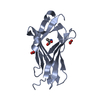
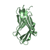
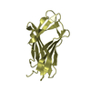
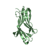
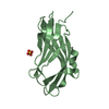
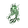
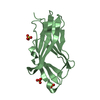
| Entry | Database: PDB / ID: 2ji0 | ||||||
|---|---|---|---|---|---|---|---|
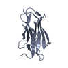
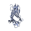
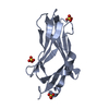
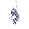
| Title | CRYSTAL STRUCTURE OF RHOGDI K138Y, K141Y MUTANT | ||||||
 Components Components | RHO GDP-DISSOCIATION INHIBITOR 1 | ||||||
 Keywords Keywords | INHIBITOR / SURFACE ENTROPY REDUCTION / GTPASE ACTIVATION / CRYSTAL ENGINEERING | ||||||
| Function / homology |  Function and homology information Function and homology informationRho GDP-dissociation inhibitor activity / Axonal growth inhibition (RHOA activation) / Axonal growth stimulation / regulation of Rho protein signal transduction / regulation of synaptic vesicle cycle / RHOC GTPase cycle / semaphorin-plexin signaling pathway / CDC42 GTPase cycle / RHOG GTPase cycle / immunological synapse ...Rho GDP-dissociation inhibitor activity / Axonal growth inhibition (RHOA activation) / Axonal growth stimulation / regulation of Rho protein signal transduction / regulation of synaptic vesicle cycle / RHOC GTPase cycle / semaphorin-plexin signaling pathway / CDC42 GTPase cycle / RHOG GTPase cycle / immunological synapse / RHOH GTPase cycle / RHOA GTPase cycle / RAC2 GTPase cycle / Rho protein signal transduction / RAC1 GTPase cycle / GTPase activator activity / Schaffer collateral - CA1 synapse / regulation of protein localization / cytoskeleton / negative regulation of apoptotic process / extracellular exosome / membrane / nucleus / cytosol Similarity search - Function | ||||||
| Biological species |  HOMO SAPIENS (human) HOMO SAPIENS (human) | ||||||
| Method |  X-RAY DIFFRACTION / MOLECULAR REPLACEMENT / Resolution: 2.1 Å X-RAY DIFFRACTION / MOLECULAR REPLACEMENT / Resolution: 2.1 Å | ||||||
 Authors Authors | Cooper, D.R. / Boczek, T. / Derewenda, Z.S. | ||||||
 Citation Citation | Journal: Acta Crystallogr.,Sect.D / Year: 2007 Title: Protein Crystallization by Surface Entropy Reduction: Optimization of the Ser Strategy Authors: Cooper, D.R. / Boczek, T. / Grelewska, K. / Pinkowska, M. / Sikorska, M. / Zawadzki, M. / Derewenda, Z.S. | ||||||
| History |
| ||||||
| Remark 700 | SHEET THE SHEET STRUCTURE OF THIS MOLECULE IS BIFURCATED. IN ORDER TO REPRESENT THIS FEATURE IN ... SHEET THE SHEET STRUCTURE OF THIS MOLECULE IS BIFURCATED. IN ORDER TO REPRESENT THIS FEATURE IN THE SHEET RECORDS BELOW, TWO SHEETS ARE DEFINED. |
- Structure visualization
| Structure viewer | Molecule: MolmilJmol/JSmol |
|---|
- Downloads & links
-Download
| PDBx/mmCIF format | 2ji0.cif.gz | 43.6 KB | Display | PDBx/mmCIF format |
|---|---|---|---|---|
| PDB format | pdb2ji0.ent.gz | 29.6 KB | Display | PDB format |
| PDBx/mmJSON format | 2ji0.json.gz | Tree view | PDBx/mmJSON format | |
| Others |  Other downloads Other downloads |
-Validation report
| Arichive directory | https://data.pdbj.org/pub/pdb/validation_reports/ji/2ji0ftp://data.pdbj.org/pub/pdb/validation_reports/ji/2ji0 | HTTPS FTP |
|---|
-Related structure data
| Related structure data |  2bxwSC 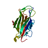 2jhsC 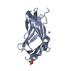 2jhtC 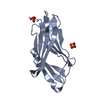 2jhuC 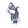 2jhvC 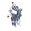 2jhwC 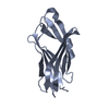 2jhxC 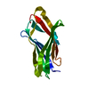 2jhyC 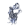 2jhzC S: Starting model for refinement C: citing same article ( |
|---|---|
| Similar structure data |


-Links
 PDBj
PDBj


- Assembly
| Deposited unit | 
| ||||||||
|---|---|---|---|---|---|---|---|---|---|
| 1 |
| ||||||||
| Unit cell |
|
-Components
| #1: Protein | Mass: 15864.039 Da / Num. of mol.: 1 / Fragment: ISOPRENYL-BINDING DOMAIN, RESIDUES 66-201 / Mutation: YES Source method: isolated from a genetically manipulated source Source: (gene. exp.) HOMO SAPIENS (human) / Production host:  | ||
|---|---|---|---|
| #2: Chemical | ChemComp-SO4 /   Mass: 96.063 Da / Num. of mol.: 1 / Source method: obtained synthetically / Formula: SO4 Mass: 96.063 Da / Num. of mol.: 1 / Source method: obtained synthetically / Formula: SO4 | ||
| #3: Water | ChemComp-HOH /  Mass: 18.015 Da / Num. of mol.: 64 / Source method: isolated from a natural source / Formula: H2O Mass: 18.015 Da / Num. of mol.: 64 / Source method: isolated from a natural source / Formula: H2O | ||
| Compound details | ENGINEERED| Sequence details | MUTATIONS ENGINEERED | |
-Experimental details
-Experiment
| Experiment | Method: X-RAY DIFFRACTION / Number of used crystals: 1 |
|---|
- Sample preparation
| Crystal | Density Matthews: 2.2 Å3/Da / Density % sol: 43.3 % |
|---|---|
| Crystal grow | pH: 7.5 Details: 32% PEG 8000, 0.22M AMMONIUM SULFATE, 0.1 M SODIUM CACODYLATE PH6.5, pH 7.50 |
-Data collection
| Diffraction | Mean temperature: 100 K |
|---|---|
| Diffraction source | Source: ROTATING ANODE / Type: ENRAF-NONIUS FR591 / Wavelength: 1.5418 |
| Detector | Type: RIGAKU IMAGE PLATE / Detector: IMAGE PLATE / Date: Sep 5, 2005 / Details: OSMIC MIRRORS |
| Radiation | Protocol: SINGLE WAVELENGTH / Monochromatic (M) / Laue (L): M / Scattering type: x-ray |
| Radiation wavelength | Wavelength: 1.5418 Å / Relative weight: 1 |
| Reflection | Resolution: 2.1→2.18 Å / Num. obs: 5179 / % possible obs: 67.5 % / Redundancy: 1.7 % / Rmerge(I) obs: 0.04 / Net I/σ(I): 18.6 |
| Reflection shell | Resolution: 2.1→2.18 Å / Redundancy: 1.4 % / Rmerge(I) obs: 0.17 / Mean I/σ(I) obs: 3.91 / % possible all: 22.5 |
- Processing
| Software |
| ||||||||||||||||||||||||||||||||||||||||||||||||||||||||||||||||||||||||||||||||||||||||||||||||||||||||||||||||||||||||||||||||||||||||||||||||||||||||||||||||||||||||||||||||||||||
|---|---|---|---|---|---|---|---|---|---|---|---|---|---|---|---|---|---|---|---|---|---|---|---|---|---|---|---|---|---|---|---|---|---|---|---|---|---|---|---|---|---|---|---|---|---|---|---|---|---|---|---|---|---|---|---|---|---|---|---|---|---|---|---|---|---|---|---|---|---|---|---|---|---|---|---|---|---|---|---|---|---|---|---|---|---|---|---|---|---|---|---|---|---|---|---|---|---|---|---|---|---|---|---|---|---|---|---|---|---|---|---|---|---|---|---|---|---|---|---|---|---|---|---|---|---|---|---|---|---|---|---|---|---|---|---|---|---|---|---|---|---|---|---|---|---|---|---|---|---|---|---|---|---|---|---|---|---|---|---|---|---|---|---|---|---|---|---|---|---|---|---|---|---|---|---|---|---|---|---|---|---|---|---|
| Refinement | Method to determine structure: MOLECULAR REPLACEMENT Starting model: PDB ENTRY 2BXW Resolution: 2.1→37.14 Å / Cor.coef. Fo:Fc: 0.958 / Cor.coef. Fo:Fc free: 0.913 / SU B: 15.95 / SU ML: 0.192 / TLS residual ADP flag: LIKELY RESIDUAL / Cross valid method: THROUGHOUT / ESU R: 1.211 / ESU R Free: 0.309 / Stereochemistry target values: MAXIMUM LIKELIHOOD / Details: HYDROGENS HAVE BEEN ADDED IN THE RIDING POSITIONS.
| ||||||||||||||||||||||||||||||||||||||||||||||||||||||||||||||||||||||||||||||||||||||||||||||||||||||||||||||||||||||||||||||||||||||||||||||||||||||||||||||||||||||||||||||||||||||
| Solvent computation | Ion probe radii: 0.8 Å / Shrinkage radii: 0.8 Å / VDW probe radii: 1.2 Å / Solvent model: MASK | ||||||||||||||||||||||||||||||||||||||||||||||||||||||||||||||||||||||||||||||||||||||||||||||||||||||||||||||||||||||||||||||||||||||||||||||||||||||||||||||||||||||||||||||||||||||
| Displacement parameters | Biso mean: 60.53 Å2
| ||||||||||||||||||||||||||||||||||||||||||||||||||||||||||||||||||||||||||||||||||||||||||||||||||||||||||||||||||||||||||||||||||||||||||||||||||||||||||||||||||||||||||||||||||||||
| Refinement step | Cycle: LAST / Resolution: 2.1→37.14 Å
| ||||||||||||||||||||||||||||||||||||||||||||||||||||||||||||||||||||||||||||||||||||||||||||||||||||||||||||||||||||||||||||||||||||||||||||||||||||||||||||||||||||||||||||||||||||||
| Refine LS restraints |
|