 Movie
Movie Controller


+ Open data

- Basic information
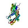
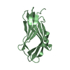
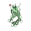
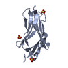
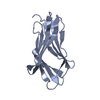
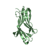
| Entry | Database: PDB / ID: 1fst | ||||||
|---|---|---|---|---|---|---|---|
| Title | CRYSTAL STRUCTURE OF TRUNCATED HUMAN RHOGDI TRIPLE MUTANT | ||||||
 Components Components | RHO GDP-DISSOCIATION INHIBITOR 1 | ||||||
 Keywords Keywords | SIGNALING PROTEIN INHIBITOR / immunoglobulin fold / beta sandwich motif / isoprenyl-binding domain / GDP-dissociation inhibitor of Rho GTPases | ||||||
| Function / homology |  Function and homology information Function and homology informationRho GDP-dissociation inhibitor activity / Axonal growth inhibition (RHOA activation) / Axonal growth stimulation / regulation of Rho protein signal transduction / regulation of synaptic vesicle cycle / RHOC GTPase cycle / semaphorin-plexin signaling pathway / CDC42 GTPase cycle / RHOG GTPase cycle / immunological synapse ...Rho GDP-dissociation inhibitor activity / Axonal growth inhibition (RHOA activation) / Axonal growth stimulation / regulation of Rho protein signal transduction / regulation of synaptic vesicle cycle / RHOC GTPase cycle / semaphorin-plexin signaling pathway / CDC42 GTPase cycle / RHOG GTPase cycle / immunological synapse / RHOH GTPase cycle / RHOA GTPase cycle / RAC2 GTPase cycle / Rho protein signal transduction / RAC1 GTPase cycle / GTPase activator activity / Schaffer collateral - CA1 synapse / regulation of protein localization / cytoskeleton / negative regulation of apoptotic process / extracellular exosome / membrane / nucleus / cytosol Similarity search - Function | ||||||
| Biological species |  Homo sapiens (human) Homo sapiens (human) | ||||||
| Method |  X-RAY DIFFRACTION / SYNCHROTRON / Resolution: 2.7 Å X-RAY DIFFRACTION / SYNCHROTRON / Resolution: 2.7 Å | ||||||
 Authors Authors | Longenecker, K.L. / Garrard, S.M. / Sheffield, P.J. / Derewenda, Z.S. | ||||||
 Citation Citation | Journal: Acta Crystallogr.,Sect.D / Year: 2001 Title: Protein crystallization by rational mutagenesis of surface residues: Lys to Ala mutations promote crystallization of RhoGDI. Authors: Longenecker, K.L. / Garrard, S.M. / Sheffield, P.J. / Derewenda, Z.S. #1: Journal: Structure / Year: 1997Title: A modulator of rho family G proteins, rhoGDI, binds these G proteins via an immunoglobulin-like domain and a flexible N-terminal arm Authors: Keep, N.H. / Barnes, M. / Barsukov, I. / Badii, R. / Lian, L. / Segal, A.W. / Moody, P.C. / Roberts, G.C. | ||||||
| History |
|
- Structure visualization
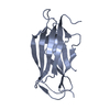
| Structure viewer | Molecule: MolmilJmol/JSmol |
|---|
- Downloads & links
-Download
| PDBx/mmCIF format | 1fst.cif.gz | 69.4 KB | Display | PDBx/mmCIF format |
|---|---|---|---|---|
| PDB format | pdb1fst.ent.gz | 52 KB | Display | PDB format |
| PDBx/mmJSON format | 1fst.json.gz | Tree view | PDBx/mmJSON format | |
| Others |  Other downloads Other downloads |
-Validation report
| Arichive directory | https://data.pdbj.org/pub/pdb/validation_reports/fs/1fstftp://data.pdbj.org/pub/pdb/validation_reports/fs/1fst | HTTPS FTP |
|---|
-Related structure data


-Links
 PDBj
PDBj


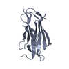
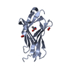
- Assembly
| Deposited unit | 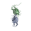
| ||||||||
|---|---|---|---|---|---|---|---|---|---|
| 1 | 
| ||||||||
| 2 | 
| ||||||||
| Unit cell |
| ||||||||
| Details | The asymmetric unit consists of two monomers. The monomer is the biologically active species. |
-Components
| #1: Protein | Mass: 20628.340 Da / Num. of mol.: 2 / Fragment: C-TERMINAL DOMAIN / Mutation: K135A, K138A, K141A Source method: isolated from a genetically manipulated source Source: (gene. exp.) Homo sapiens (human) / Plasmid: PGEX / Production host:  #2: Water | ChemComp-HOH / |  Mass: 18.015 Da / Num. of mol.: 40 / Source method: isolated from a natural source / Formula: H2O Mass: 18.015 Da / Num. of mol.: 40 / Source method: isolated from a natural source / Formula: H2O |
|---|
-Experimental details
-Experiment
| Experiment | Method: X-RAY DIFFRACTION / Number of used crystals: 1 |
|---|
- Sample preparation
| Crystal | Density Matthews: 3.02 Å3/Da / Density % sol: 59.29 % | |||||||||||||||||||||||||
|---|---|---|---|---|---|---|---|---|---|---|---|---|---|---|---|---|---|---|---|---|---|---|---|---|---|---|
| Crystal grow | Temperature: 293 K / Method: vapor diffusion, hanging drop / pH: 7 Details: PEG 3400, isopropanol, HEPES buffer, pH 7.0, VAPOR DIFFUSION, HANGING DROP, temperature 293.0K | |||||||||||||||||||||||||
| Crystal grow | *PLUS Method: vapor diffusion | |||||||||||||||||||||||||
| Components of the solutions | *PLUS
|
-Data collection
| Diffraction | Mean temperature: 100 K |
|---|---|
| Diffraction source | Source: SYNCHROTRON / Site: EMBL/DESY, HAMBURG  / Beamline: X11 / Wavelength: 0.9091 / Beamline: X11 / Wavelength: 0.9091 |
| Detector | Type: MAR CCD 165 mm / Detector: CCD / Date: Dec 6, 1999 |
| Radiation | Protocol: SINGLE WAVELENGTH / Monochromatic (M) / Laue (L): M / Scattering type: x-ray |
| Radiation wavelength | Wavelength: 0.9091 Å / Relative weight: 1 |
| Reflection | Resolution: 2.7→20 Å / Num. all: 13241 / Num. obs: 13241 / % possible obs: 99.6 % / Observed criterion σ(F): 0 / Observed criterion σ(I): 0 / Redundancy: 3.8 % / Biso Wilson estimate: 73 Å2 / Rmerge(I) obs: 0.048 / Net I/σ(I): 17.9 |
| Reflection shell | Resolution: 2.7→2.78 Å / Redundancy: 3.8 % / Rmerge(I) obs: 0.267 / Num. unique all: 1129 / % possible all: 99.6 |
| Reflection | *PLUS Num. measured all: 49713 |
| Reflection shell | *PLUS % possible obs: 99.6 % / Mean I/σ(I) obs: 3.7 |
- Processing
| Software |
| |||||||||||||||||||||||||
|---|---|---|---|---|---|---|---|---|---|---|---|---|---|---|---|---|---|---|---|---|---|---|---|---|---|---|
| Refinement | Resolution: 2.7→20 Å / σ(F): 0 / σ(I): 0 / Stereochemistry target values: Engh and Huber
| |||||||||||||||||||||||||
| Refinement step | Cycle: LAST / Resolution: 2.7→20 Å
| |||||||||||||||||||||||||
| Refine LS restraints |
| |||||||||||||||||||||||||
| Software | *PLUS Name: CNS / Version: 0.9 / Classification: refinement | |||||||||||||||||||||||||
| Refine LS restraints | *PLUS
|
