 Movie
Movie Controller

[English] 日本語
 Yorodumi
- PDB-6hyc: The structure of full-length human phenylalanine hydroxylase in c... -
+ Open data

- Basic information
| Entry | Database: PDB / ID: 6hyc | |||||||||
|---|---|---|---|---|---|---|---|---|---|---|
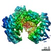
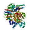
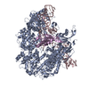
| Title | The structure of full-length human phenylalanine hydroxylase in complex with the cofactor and negative regulator tetrahydrobiopterin | |||||||||
 Components Components | Phenylalanine-4-hydroxylase | |||||||||
 Keywords Keywords | OXIDOREDUCTASE / Tetrahydrobiopterin / phenylalanine hydroxylase / phenylketonuria / allostery | |||||||||
| Function / homology |  Function and homology information Function and homology informationPhenylketonuria / phenylalanine 4-monooxygenase / Phenylalanine metabolism / phenylalanine 4-monooxygenase activity / L-tyrosine biosynthetic process / catecholamine biosynthetic process / L-phenylalanine catabolic process / amino acid biosynthetic process / iron ion binding / cytosol Similarity search - Function | |||||||||
| Biological species |  Homo sapiens (human) Homo sapiens (human) | |||||||||
| Method |  X-RAY DIFFRACTION / SYNCHROTRON / MOLECULAR REPLACEMENT / Resolution: 3.18 Å X-RAY DIFFRACTION / SYNCHROTRON / MOLECULAR REPLACEMENT / Resolution: 3.18 Å | |||||||||
 Authors Authors | Alcorlo Pages, M. / Flydal, I.M. | |||||||||
| Funding support |  Spain, Spain,  Norway, 2items Norway, 2items
| |||||||||
 Citation Citation | Journal: Proc Natl Acad Sci U S A / Year: 2019 Title: Structure of full-length human phenylalanine hydroxylase in complex with tetrahydrobiopterin. Authors: Marte Innselset Flydal / Martín Alcorlo-Pagés / Fredrik Gullaksen Johannessen / Siseth Martínez-Caballero / Lars Skjærven / Rafael Fernandez-Leiro / Aurora Martinez / Juan A Hermoso / Abstract: Phenylalanine hydroxylase (PAH) is a key enzyme in the catabolism of phenylalanine, and mutations in this enzyme cause phenylketonuria (PKU), a genetic disorder that leads to brain damage and mental ...Phenylalanine hydroxylase (PAH) is a key enzyme in the catabolism of phenylalanine, and mutations in this enzyme cause phenylketonuria (PKU), a genetic disorder that leads to brain damage and mental retardation if untreated. Some patients benefit from supplementation with a synthetic formulation of the cofactor tetrahydrobiopterin (BH) that partly acts as a pharmacological chaperone. Here we present structures of full-length human PAH (hPAH) both unbound and complexed with BH in the precatalytic state. Crystal structures, solved at 3.18-Å resolution, show the interactions between the cofactor and PAH, explaining the negative regulation exerted by BH BH forms several H-bonds with the N-terminal autoregulatory tail but is far from the catalytic Fe Upon BH binding a polar and salt-bridge interaction network links the three PAH domains, explaining the stability conferred by BH Importantly, BH binding modulates the interaction between subunits, providing information about PAH allostery. Moreover, we also show that the cryo-EM structure of hPAH in absence of BH reveals a highly dynamic conformation for the tetramers. Structural analyses of the hPAH:BH subunits revealed that the substrate-induced movement of Tyr138 into the active site could be coupled to the displacement of BH from the precatalytic toward the active conformation, a molecular mechanism that was supported by site-directed mutagenesis and targeted molecular dynamics simulations. Finally, comparison of the rat and human PAH structures show that hPAH is more dynamic, which is related to amino acid substitutions that enhance the flexibility of hPAH and may increase the susceptibility to PKU-associated mutations. | |||||||||
| History |
|
- Structure visualization
| Structure viewer | Molecule: MolmilJmol/JSmol |
|---|
- Downloads & links
-Download
| PDBx/mmCIF format | 6hyc.cif.gz | 348 KB | Display | PDBx/mmCIF format |
|---|---|---|---|---|
| PDB format | pdb6hyc.ent.gz | 284.3 KB | Display | PDB format |
| PDBx/mmJSON format | 6hyc.json.gz | Tree view | PDBx/mmJSON format | |
| Others |  Other downloads Other downloads |
-Validation report
| Arichive directory | https://data.pdbj.org/pub/pdb/validation_reports/hy/6hycftp://data.pdbj.org/pub/pdb/validation_reports/hy/6hyc | HTTPS FTP |
|---|
-Related structure data
| Related structure data |  4605C  6hpoC  5denS S: Starting model for refinement C: citing same article ( |
|---|---|
| Similar structure data |

-Links
 PDBj
PDBj


- Assembly
| Deposited unit | 
| ||||||||
|---|---|---|---|---|---|---|---|---|---|
| 1 | 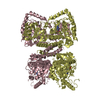
| ||||||||
| 2 | 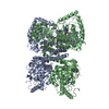
| ||||||||
| Unit cell |
|
-Components
| #1: Protein | Mass: 51928.906 Da / Num. of mol.: 4 Source method: isolated from a genetically manipulated source Source: (gene. exp.) Homo sapiens (human) / Gene: PAH / Production host:  #2: Chemical |   Mass: 241.247 Da / Num. of mol.: 3 / Source method: obtained synthetically / Formula: C9H15N5O3 Mass: 241.247 Da / Num. of mol.: 3 / Source method: obtained synthetically / Formula: C9H15N5O3#3: Water | ChemComp-HOH / |  Mass: 18.015 Da / Num. of mol.: 5 / Source method: isolated from a natural source / Formula: H2O Mass: 18.015 Da / Num. of mol.: 5 / Source method: isolated from a natural source / Formula: H2O |
|---|
-Experimental details
-Experiment
| Experiment | Method: X-RAY DIFFRACTION / Number of used crystals: 1 |
|---|
- Sample preparation
| Crystal | Density Matthews: 2.48 Å3/Da / Density % sol: 50.35 % |
|---|---|
| Crystal grow | Temperature: 291 K / Method: vapor diffusion, sitting drop / pH: 7 Details: 1.5 M DL Malic Acid pH=7.0, 100 mM Bis-Tris Propane pH=6.2, 0.9 mM Thesit, 0.98 mM BH4, 25 mM DTT and 1 mM reduced glutathione |
-Data collection
| Diffraction | Mean temperature: 100 K / Serial crystal experiment: N |
|---|---|
| Diffraction source | Source: SYNCHROTRON / Site: ALBA / Beamline: XALOC / Wavelength: 0.97926 Å |
| Detector | Type: DECTRIS PILATUS3 S 6M / Detector: PIXEL / Date: Oct 19, 2016 |
| Radiation | Protocol: SINGLE WAVELENGTH / Monochromatic (M) / Laue (L): M / Scattering type: x-ray |
| Radiation wavelength | Wavelength: 0.97926 Å / Relative weight: 1 |
| Reflection | Resolution: 3.18→33.92 Å / Num. obs: 34280 / % possible obs: 94.3 % / Redundancy: 3 % / Rmerge(I) obs: 0.181 / Rpim(I) all: 0.1314 / Net I/σ(I): 2.4 |
| Reflection shell | Resolution: 3.18→3.34 Å / Num. unique obs: 3926 |
- Processing
| Software |
| ||||||||||||||||||||||||||||||||||||||||||||||||||||||||||||||||||||||||||||||||||||
|---|---|---|---|---|---|---|---|---|---|---|---|---|---|---|---|---|---|---|---|---|---|---|---|---|---|---|---|---|---|---|---|---|---|---|---|---|---|---|---|---|---|---|---|---|---|---|---|---|---|---|---|---|---|---|---|---|---|---|---|---|---|---|---|---|---|---|---|---|---|---|---|---|---|---|---|---|---|---|---|---|---|---|---|---|---|
| Refinement | Method to determine structure: MOLECULAR REPLACEMENT Starting model: 5den Resolution: 3.18→33.92 Å / SU ML: 0.43 / Cross valid method: FREE R-VALUE / σ(F): 0 / Phase error: 31.58
| ||||||||||||||||||||||||||||||||||||||||||||||||||||||||||||||||||||||||||||||||||||
| Solvent computation | Shrinkage radii: 0.9 Å / VDW probe radii: 1.11 Å | ||||||||||||||||||||||||||||||||||||||||||||||||||||||||||||||||||||||||||||||||||||
| Refinement step | Cycle: LAST / Resolution: 3.18→33.92 Å
| ||||||||||||||||||||||||||||||||||||||||||||||||||||||||||||||||||||||||||||||||||||
| Refine LS restraints |
| ||||||||||||||||||||||||||||||||||||||||||||||||||||||||||||||||||||||||||||||||||||
| LS refinement shell |
|
