 Movie
Movie Controller

[English] 日本語
 Yorodumi
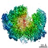
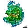
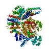
- PDB-5egq: Structure of tetrameric rat phenylalanine hydroxylase mutant R270... -
+ Open data

- Basic information
| Entry | Database: PDB / ID: 5egq | ||||||
|---|---|---|---|---|---|---|---|
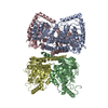
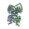
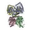
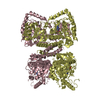
| Title | Structure of tetrameric rat phenylalanine hydroxylase mutant R270K, residues 25-453 | ||||||
 Components Components | Phenylalanine-4-hydroxylase | ||||||
 Keywords Keywords | OXIDOREDUCTASE / hydroxylase / phenylketonuria / PKU mutation / allostery / ACT domain | ||||||
| Function / homology |  Function and homology information Function and homology information: / Phenylalanine metabolism / : / phenylalanine 4-monooxygenase / phenylalanine 4-monooxygenase activity / L-tyrosine biosynthetic process / L-phenylalanine catabolic process / amino acid binding / iron ion binding / identical protein binding Similarity search - Function | ||||||
| Biological species |  | ||||||
| Method |  X-RAY DIFFRACTION / SYNCHROTRON / MOLECULAR REPLACEMENT / Resolution: 2.5 Å X-RAY DIFFRACTION / SYNCHROTRON / MOLECULAR REPLACEMENT / Resolution: 2.5 Å | ||||||
 Authors Authors | Taylor, A.B. / Khan, C.A. / Fitzpatrick, P.F. | ||||||
 Citation Citation | Journal: J.Am.Chem.Soc. / Year: 2016 Title: Domain Movements upon Activation of Phenylalanine Hydroxylase Characterized by Crystallography and Chromatography-Coupled Small-Angle X-ray Scattering. Authors: Meisburger, S.P. / Taylor, A.B. / Khan, C.A. / Zhang, S. / Fitzpatrick, P.F. / Ando, N. | ||||||
| History |
|
- Structure visualization
| Structure viewer | Molecule: MolmilJmol/JSmol |
|---|
- Downloads & links
-Download
| PDBx/mmCIF format | 5egq.cif.gz | 333.9 KB | Display | PDBx/mmCIF format |
|---|---|---|---|---|
| PDB format | pdb5egq.ent.gz | 272 KB | Display | PDB format |
| PDBx/mmJSON format | 5egq.json.gz | Tree view | PDBx/mmJSON format | |
| Others |  Other downloads Other downloads |
-Validation report
| Arichive directory | https://data.pdbj.org/pub/pdb/validation_reports/eg/5egqftp://data.pdbj.org/pub/pdb/validation_reports/eg/5egq | HTTPS FTP |
|---|
-Related structure data
| Related structure data |  5fgjC  2phmS S: Starting model for refinement C: citing same article ( |
|---|---|
| Similar structure data |

-Links
 PDBj
PDBj


- Assembly
| Deposited unit | 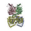
| ||||||||
|---|---|---|---|---|---|---|---|---|---|
| 1 |
| ||||||||
| Unit cell |
|
-Components
| #1: Protein | Mass: 51857.566 Da / Num. of mol.: 4 / Mutation: R270K Source method: isolated from a genetically manipulated source Source: (gene. exp.)  #2: Chemical | ChemComp-SO4 /   Mass: 96.063 Da / Num. of mol.: 6 / Source method: obtained synthetically / Formula: SO4 Mass: 96.063 Da / Num. of mol.: 6 / Source method: obtained synthetically / Formula: SO4#3: Water | ChemComp-HOH / |  Mass: 18.015 Da / Num. of mol.: 176 / Source method: isolated from a natural source / Formula: H2O Mass: 18.015 Da / Num. of mol.: 176 / Source method: isolated from a natural source / Formula: H2O |
|---|
-Experimental details
-Experiment
| Experiment | Method: X-RAY DIFFRACTION / Number of used crystals: 1 |
|---|
- Sample preparation
| Crystal | Density Matthews: 2.28 Å3/Da / Density % sol: 45.98 % |
|---|---|
| Crystal grow | Temperature: 295 K / Method: vapor diffusion, sitting drop / pH: 6.5 Details: 0.1 M bis-tris:HCl, 25% PEG 3350, 0.2 M ammonium sulfate |
-Data collection
| Diffraction | Mean temperature: 100 K |
|---|---|
| Diffraction source | Source: SYNCHROTRON / Site: APS  / Beamline: 24-ID-C / Wavelength: 0.9791 Å / Beamline: 24-ID-C / Wavelength: 0.9791 Å |
| Detector | Type: PSI PILATUS 6M / Detector: PIXEL / Date: Apr 22, 2015 |
| Radiation | Protocol: SINGLE WAVELENGTH / Monochromatic (M) / Laue (L): M / Scattering type: x-ray |
| Radiation wavelength | Wavelength: 0.9791 Å / Relative weight: 1 |
| Reflection | Resolution: 2.5→47.16 Å / Num. obs: 61203 / % possible obs: 99 % / Redundancy: 3.7 % / Biso Wilson estimate: 38.6 Å2 / Rsym value: 0.096 / Net I/σ(I): 10.1 |
| Reflection shell | Resolution: 2.5→2.64 Å / Redundancy: 3.8 % / Rmerge(I) obs: 0.623 / Mean I/σ(I) obs: 2 / % possible all: 99.7 |
- Processing
| Software |
| |||||||||||||||||||||||||||||||||||||||||||||||||||||||||||||||||||||||||||||||||||||||||||||||||||||||||
|---|---|---|---|---|---|---|---|---|---|---|---|---|---|---|---|---|---|---|---|---|---|---|---|---|---|---|---|---|---|---|---|---|---|---|---|---|---|---|---|---|---|---|---|---|---|---|---|---|---|---|---|---|---|---|---|---|---|---|---|---|---|---|---|---|---|---|---|---|---|---|---|---|---|---|---|---|---|---|---|---|---|---|---|---|---|---|---|---|---|---|---|---|---|---|---|---|---|---|---|---|---|---|---|---|---|---|
| Refinement | Method to determine structure: MOLECULAR REPLACEMENT Starting model: 2PHM Resolution: 2.5→47.155 Å / SU ML: 0.34 / Cross valid method: THROUGHOUT / σ(F): 1.35 / Phase error: 28.61 / Stereochemistry target values: ML
| |||||||||||||||||||||||||||||||||||||||||||||||||||||||||||||||||||||||||||||||||||||||||||||||||||||||||
| Solvent computation | Shrinkage radii: 0.9 Å / VDW probe radii: 1.11 Å / Solvent model: FLAT BULK SOLVENT MODEL | |||||||||||||||||||||||||||||||||||||||||||||||||||||||||||||||||||||||||||||||||||||||||||||||||||||||||
| Refinement step | Cycle: LAST / Resolution: 2.5→47.155 Å
| |||||||||||||||||||||||||||||||||||||||||||||||||||||||||||||||||||||||||||||||||||||||||||||||||||||||||
| Refine LS restraints |
| |||||||||||||||||||||||||||||||||||||||||||||||||||||||||||||||||||||||||||||||||||||||||||||||||||||||||
| LS refinement shell |
|
