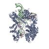
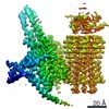
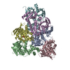
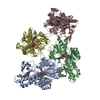
Journal: Proc Natl Acad Sci U S A / Year: 2019 Title: Structure of full-length human phenylalanine hydroxylase in complex with tetrahydrobiopterin. Authors: Marte Innselset Flydal / Martín Alcorlo-Pagés / Fredrik Gullaksen Johannessen / Siseth Martínez-Caballero / Lars Skjærven / Rafael Fernandez-Leiro / Aurora Martinez / Juan A Hermoso / Abstract: Phenylalanine hydroxylase (PAH) is a key enzyme in the catabolism of phenylalanine, and mutations in this enzyme cause phenylketonuria (PKU), a genetic disorder that leads to brain damage and mental ...Phenylalanine hydroxylase (PAH) is a key enzyme in the catabolism of phenylalanine, and mutations in this enzyme cause phenylketonuria (PKU), a genetic disorder that leads to brain damage and mental retardation if untreated. Some patients benefit from supplementation with a synthetic formulation of the cofactor tetrahydrobiopterin (BH) that partly acts as a pharmacological chaperone. Here we present structures of full-length human PAH (hPAH) both unbound and complexed with BH in the precatalytic state. Crystal structures, solved at 3.18-Å resolution, show the interactions between the cofactor and PAH, explaining the negative regulation exerted by BH BH forms several H-bonds with the N-terminal autoregulatory tail but is far from the catalytic Fe Upon BH binding a polar and salt-bridge interaction network links the three PAH domains, explaining the stability conferred by BH Importantly, BH binding modulates the interaction between subunits, providing information about PAH allostery. Moreover, we also show that the cryo-EM structure of hPAH in absence of BH reveals a highly dynamic conformation for the tetramers. Structural analyses of the hPAH:BH subunits revealed that the substrate-induced movement of Tyr138 into the active site could be coupled to the displacement of BH from the precatalytic toward the active conformation, a molecular mechanism that was supported by site-directed mutagenesis and targeted molecular dynamics simulations. Finally, comparison of the rat and human PAH structures show that hPAH is more dynamic, which is related to amino acid substitutions that enhance the flexibility of hPAH and may increase the susceptibility to PKU-associated mutations.
History
Deposition
Feb 11, 2019
-
Header (metadata) release
Feb 27, 2019
-
Map release
Jun 5, 2019
-
Update
Nov 25, 2020
-
Current status
Nov 25, 2020
Processing site: PDBe / Status: Released
-
Structure visualization
Movie
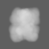
Surface view with section colored by density value
Cryogen name: ETHANE / Chamber humidity: 95 % / Chamber temperature: 277 K / Instrument: FEI VITROBOT MARK III Details: 3microL, no incubation, 2 seconds blot, no wait before plunging.
Details
The thawed sample was run through SEC (S200I) prior to grid preparation
-
Electron microscopy
Microscope
FEI TITAN KRIOS
Specialist optics
Energy filter - Name: GIF Quantum LS / Energy filter - Slit width: 20 eV
Image recording
Film or detector model: GATAN K2 SUMMIT (4k x 4k) / Detector mode: COUNTING / Digitization - Dimensions - Width: 3838 pixel / Digitization - Dimensions - Height: 3710 pixel / Digitization - Frames/image: 1-80 / Number grids imaged: 1 / Number real images: 1493 / Average exposure time: 12.0 sec. / Average electron dose: 42.0 e/Å2
Electron beam
Acceleration voltage: 300 kV / Electron source: FIELD EMISSION GUN
In the structure databanks used in Yorodumi, some data are registered as the other names, "COVID-19 virus" and "2019-nCoV". Here are the details of the virus and the list of structure data.
Jan 31, 2019. EMDB accession codes are about to change! (news from PDBe EMDB page)
EMDB accession codes are about to change! (news from PDBe EMDB page)
The allocation of 4 digits for EMDB accession codes will soon come to an end. Whilst these codes will remain in use, new EMDB accession codes will include an additional digit and will expand incrementally as the available range of codes is exhausted. The current 4-digit format prefixed with “EMD-” (i.e. EMD-XXXX) will advance to a 5-digit format (i.e. EMD-XXXXX), and so on. It is currently estimated that the 4-digit codes will be depleted around Spring 2019, at which point the 5-digit format will come into force.
The EM Navigator/Yorodumi systems omit the EMD- prefix.
Related info.:Q: What is EMD? / ID/Accession-code notation in Yorodumi/EM Navigator
Yorodumi is a browser for structure data from EMDB, PDB, SASBDB, etc.
This page is also the successor to EM Navigator detail page, and also detail information page/front-end page for Omokage search.
The word "yorodu" (or yorozu) is an old Japanese word meaning "ten thousand". "mi" (miru) is to see.
Related info.:EMDB / PDB / SASBDB / Comparison of 3 databanks / Yorodumi Search / Aug 31, 2016. New EM Navigator & Yorodumi / Yorodumi Papers / Jmol/JSmol / Function and homology information / Changes in new EM Navigator and Yorodumi
 Movie
Movie Controller


 Open data
 Basic information
 Map data
 Sample
 Function and homology information
 Homo sapiens (human)
 Authors
 Spain, 1 items
  Citation
 Structure visualization
 Downloads & links
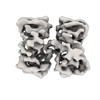 emd_4605.png
 http://ftp.pdbj.org/pub/emdb/structures/EMD-4605


 Z
Z Y
Y X
X


 Sample components
 Processing
 Electron microscopy
 FIELD EMISSION GUN