[English] 日本語
Yorodumi
- PDB-6hpo: Crystallographic structure of the catalytic domain of Human Pheny... -

+
Open data


ID or keywords:

Loading...

-
Basic information

Entry
Database: PDB / ID: 6hpo
TitleCrystallographic structure of the catalytic domain of Human Phenylalanine Hydroxylase (hPAH CD) in complex with iron at 1.6 Angstrom
ComponentsPhenylalanine-4-hydroxylase
KeywordsOXIDOREDUCTASE / Tetrahydrobiopterin / amino acid hydroxylases / phenylketonuria / allosteric regulation / METAL BINDING PROTEIN
Function / homology
Function and homology information


Phenylketonuria / Phenylalanine metabolism / phenylalanine 4-monooxygenase / phenylalanine 4-monooxygenase activity / tyrosine biosynthetic process / catecholamine biosynthetic process / L-phenylalanine catabolic process / amino acid biosynthetic process / iron ion binding / cytosol
Similarity search - Function
Phenylalanine-4-hydroxylase, tetrameric form / Eukaryotic phenylalanine-4-hydroxylase, catalytic domain / Phenylalanine Hydroxylase / Aromatic amino acid hydroxylase / Tyrosine 3-monooxygenase-like / ACT domain / Aromatic amino acid hydroxylase, iron/copper binding site / Biopterin-dependent aromatic amino acid hydroxylases signature. / Aromatic amino acid hydroxylase / Aromatic amino acid hydroxylase, C-terminal ...Phenylalanine-4-hydroxylase, tetrameric form / Eukaryotic phenylalanine-4-hydroxylase, catalytic domain / Phenylalanine Hydroxylase / Aromatic amino acid hydroxylase / Tyrosine 3-monooxygenase-like / ACT domain / Aromatic amino acid hydroxylase, iron/copper binding site / Biopterin-dependent aromatic amino acid hydroxylases signature. / Aromatic amino acid hydroxylase / Aromatic amino acid hydroxylase, C-terminal / Aromatic amino acid monoxygenase, C-terminal domain superfamily / Aromatic amino acid hydroxylase superfamily / Biopterin-dependent aromatic amino acid hydroxylase / Biopterin-dependent aromatic amino acid hydroxylase family profile. / ACT domain profile. / ACT domain / ACT-like domain / Orthogonal Bundle / Mainly Alpha
Similarity search - Domain/homology
: / Phenylalanine-4-hydroxylase
Similarity search - Component
Biological speciesHomo sapiens (human)
MethodX-RAY DIFFRACTION / SYNCHROTRON / MOLECULAR REPLACEMENT / Resolution: 1.67 Å
AuthorsAlcorlo Pages, M. / Innselset Flydal, M.
Funding support Spain, Norway, 2items
OrganizationGrant numberCountry
Spanish Ministry of Economy and CompetitivenessBFU2014-59389-P and BFU2017-90030-P to JAH Spain
Research Council of NorwayPrograms Forny (248889/O30) and FRIMEDBIO (261826) to AM Norway
Citation
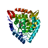
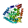
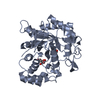
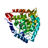
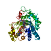
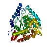
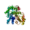
Journal: Proc Natl Acad Sci U S A / Year: 2019
Title: Structure of full-length human phenylalanine hydroxylase in complex with tetrahydrobiopterin.
Authors: Marte Innselset Flydal / Martín Alcorlo-Pagés / Fredrik Gullaksen Johannessen / Siseth Martínez-Caballero / Lars Skjærven / Rafael Fernandez-Leiro / Aurora Martinez / Juan A Hermoso /
Abstract: Phenylalanine hydroxylase (PAH) is a key enzyme in the catabolism of phenylalanine, and mutations in this enzyme cause phenylketonuria (PKU), a genetic disorder that leads to brain damage and mental ...Phenylalanine hydroxylase (PAH) is a key enzyme in the catabolism of phenylalanine, and mutations in this enzyme cause phenylketonuria (PKU), a genetic disorder that leads to brain damage and mental retardation if untreated. Some patients benefit from supplementation with a synthetic formulation of the cofactor tetrahydrobiopterin (BH) that partly acts as a pharmacological chaperone. Here we present structures of full-length human PAH (hPAH) both unbound and complexed with BH in the precatalytic state. Crystal structures, solved at 3.18-Å resolution, show the interactions between the cofactor and PAH, explaining the negative regulation exerted by BH BH forms several H-bonds with the N-terminal autoregulatory tail but is far from the catalytic Fe Upon BH binding a polar and salt-bridge interaction network links the three PAH domains, explaining the stability conferred by BH Importantly, BH binding modulates the interaction between subunits, providing information about PAH allostery. Moreover, we also show that the cryo-EM structure of hPAH in absence of BH reveals a highly dynamic conformation for the tetramers. Structural analyses of the hPAH:BH subunits revealed that the substrate-induced movement of Tyr138 into the active site could be coupled to the displacement of BH from the precatalytic toward the active conformation, a molecular mechanism that was supported by site-directed mutagenesis and targeted molecular dynamics simulations. Finally, comparison of the rat and human PAH structures show that hPAH is more dynamic, which is related to amino acid substitutions that enhance the flexibility of hPAH and may increase the susceptibility to PKU-associated mutations.
History
DepositionSep 21, 2018Deposition site: PDBE / Processing site: PDBE
Revision 1.0Jun 5, 2019Provider: repository / Type: Initial release
Revision 1.1Jun 19, 2019Group: Data collection / Database references / Category: citation / citation_author / pdbx_database_proc
Item: _citation.journal_volume / _citation.page_first ..._citation.journal_volume / _citation.page_first / _citation.page_last / _citation_author.identifier_ORCID
Revision 1.2Jan 24, 2024Group: Data collection / Database references ...Data collection / Database references / Derived calculations / Refinement description
Category: chem_comp_atom / chem_comp_bond ...chem_comp_atom / chem_comp_bond / database_2 / pdbx_initial_refinement_model / pdbx_struct_conn_angle / struct_conn
Item: _database_2.pdbx_DOI / _database_2.pdbx_database_accession ..._database_2.pdbx_DOI / _database_2.pdbx_database_accession / _pdbx_struct_conn_angle.ptnr1_auth_seq_id / _pdbx_struct_conn_angle.ptnr3_auth_seq_id / _pdbx_struct_conn_angle.value / _struct_conn.pdbx_dist_value / _struct_conn.ptnr2_auth_seq_id

-
Structure visualization

Structure viewerMolecule:
MolmilJmol/JSmol

Downloads & links

-
Assembly

Deposited unit
A: Phenylalanine-4-hydroxylase
hetero molecules


Theoretical massNumber of molelcules
Total (without water)51,9852
Polymers51,9291
Non-polymers561
Water5,296294
1


  • Idetical with deposited unit
  • defined by author&software
TypeNameSymmetry operationNumber
identity operation1_555x,y,z1
Buried area140 Å2
ΔGint-13 kcal/mol
Surface area14520 Å2
MethodPISA
Unit cell
Length a, b, c (Å)65.856, 107.549, 124.016
Angle α, β, γ (deg.)90.00, 90.00, 90.00
Int Tables number20
Space group name H-MC2221

-
Components

#1: Protein Phenylalanine-4-hydroxylase / PAH / Phe-4-monooxygenase


Mass: 51928.906 Da / Num. of mol.: 1
Source method: isolated from a genetically manipulated source
Source: (gene. exp.) Homo sapiens (human) / Gene: PAH / Production host: Escherichia coli (E. coli) / References: UniProt: P00439, phenylalanine 4-monooxygenase
#2: Chemical ChemComp-FE / FE (III) ION / Iron


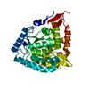
Mass: 55.845 Da / Num. of mol.: 1 / Source method: obtained synthetically / Formula: Fe
#3: Water ChemComp-HOH / water / Water


Mass: 18.015 Da / Num. of mol.: 294 / Source method: isolated from a natural source / Formula: H2O

-
Experimental details

-
Experiment

ExperimentMethod: X-RAY DIFFRACTION / Number of used crystals: 1

-
Sample preparation

CrystalDensity Matthews: 2.48 Å3/Da / Density % sol: 50.35 %
Crystal growTemperature: 291 K / Method: vapor diffusion / Details: 40 mM PIPES, 20% PEG 2000, pH 6.8

-
Data collection

DiffractionMean temperature: 100 K / Serial crystal experiment: N
Diffraction sourceSource: SYNCHROTRON / Site: ALBA / Beamline: XALOC / Wavelength: 0.9792 Å
DetectorType: DECTRIS PILATUS3 6M / Detector: PIXEL / Date: Mar 1, 2018
RadiationProtocol: SINGLE WAVELENGTH / Monochromatic (M) / Laue (L): M / Scattering type: x-ray
Radiation wavelengthWavelength: 0.9792 Å / Relative weight: 1
ReflectionResolution: 1.67→32.77 Å / Num. obs: 51389 / % possible obs: 99.96 % / Redundancy: 13.4 % / Rmerge(I) obs: 0.084 / Rpim(I) all: 0.026 / Net I/σ(I): 16.2
Reflection shellResolution: 1.67→1.73 Å / Rmerge(I) obs: 1.921 / Num. unique obs: 2603 / Rpim(I) all: 0.574

-
Processing

Software
NameVersionClassification
PHENIX(1.13_2998: ???)refinement
XDSdata reduction
Aimlessdata scaling
MOLREPphasing
RefinementMethod to determine structure: MOLECULAR REPLACEMENT
Starting model: 4ANP
Resolution: 1.67→32.77 Å / SU ML: 0.16 / Cross valid method: FREE R-VALUE / σ(F): 1.33 / Phase error: 19.41
RfactorNum. reflection% reflection
Rfree0.177 2541 4.95 %
Rwork0.1607 --
obs0.1615 51335 99.92 %
Solvent computationShrinkage radii: 0.9 Å / VDW probe radii: 1.11 Å
Refinement stepCycle: LAST / Resolution: 1.67→32.77 Å
ProteinNucleic acidLigandSolventTotal
Num. atoms2540 0 1 294 2835
Refine LS restraints
Refine-IDTypeDev idealNumber
X-RAY DIFFRACTIONf_bond_d0.0062680
X-RAY DIFFRACTIONf_angle_d0.7733651
X-RAY DIFFRACTIONf_dihedral_angle_d5.8822208
X-RAY DIFFRACTIONf_chiral_restr0.05382
X-RAY DIFFRACTIONf_plane_restr0.005476
LS refinement shell
Resolution (Å)Rfactor RfreeNum. reflection RfreeRfactor RworkNum. reflection RworkRefine-ID% reflection obs (%)
1.67-1.70210.30071450.27322706X-RAY DIFFRACTION100
1.7021-1.73690.28131310.25322671X-RAY DIFFRACTION100
1.7369-1.77460.23471360.2252656X-RAY DIFFRACTION100
1.7746-1.81590.22341320.21072696X-RAY DIFFRACTION100
1.8159-1.86130.21071130.19992718X-RAY DIFFRACTION100
1.8613-1.91160.20941460.18822681X-RAY DIFFRACTION100
1.9116-1.96790.20441270.1672706X-RAY DIFFRACTION100
1.9679-2.03140.1791400.15962681X-RAY DIFFRACTION100
2.0314-2.1040.17261610.15852687X-RAY DIFFRACTION100
2.104-2.18820.18221520.16262660X-RAY DIFFRACTION100
2.1882-2.28780.19891410.15832709X-RAY DIFFRACTION100
2.2878-2.40840.18331490.15962680X-RAY DIFFRACTION100
2.4084-2.55920.17731440.15642721X-RAY DIFFRACTION100
2.5592-2.75670.20381450.16512726X-RAY DIFFRACTION100
2.7567-3.0340.18211530.1632708X-RAY DIFFRACTION100
3.034-3.47260.17671330.15682754X-RAY DIFFRACTION100
3.4726-4.37340.16011590.13662739X-RAY DIFFRACTION100
4.3734-32.93440.14021340.15452895X-RAY DIFFRACTION100
Refinement TLS params.Method: refined / Origin x: 0.1353 Å / Origin y: 24.8754 Å / Origin z: 9.606 Å
111213212223313233
T0.2107 Å20.0059 Å20.0035 Å2-0.2229 Å2-0.0025 Å2--0.2037 Å2
L1.071 °2-0.209 °2-0.1755 °2-0.7995 °20.1815 °2--0.6804 °2
S-0.0217 Å °-0.0939 Å °-0.0357 Å °-0.0221 Å °0.0171 Å °-0.0176 Å °-0.0102 Å °-0.0493 Å °0.0063 Å °
Refinement TLS groupSelection details: all

+
About Yorodumi

-
News

-
Feb 9, 2022. New format data for meta-information of EMDB entries

New format data for meta-information of EMDB entries

  • Version 3 of the EMDB header file is now the official format.
  • The previous official version 1.9 will be removed from the archive.

Related info.:EMDB header

External links:wwPDB to switch to version 3 of the EMDB data model

-
Aug 12, 2020. Covid-19 info

Covid-19 info

URL: https://pdbj.org/emnavi/covid19.php

New page: Covid-19 featured information page in EM Navigator.

Related info.:Covid-19 info / Mar 5, 2020. Novel coronavirus structure data

+
Mar 5, 2020. Novel coronavirus structure data

Novel coronavirus structure data

Related info.:Yorodumi Speices / Aug 12, 2020. Covid-19 info

External links:COVID-19 featured content - PDBj / Molecule of the Month (242):Coronavirus Proteases

+
Jan 31, 2019. EMDB accession codes are about to change! (news from PDBe EMDB page)

EMDB accession codes are about to change! (news from PDBe EMDB page)

  • The allocation of 4 digits for EMDB accession codes will soon come to an end. Whilst these codes will remain in use, new EMDB accession codes will include an additional digit and will expand incrementally as the available range of codes is exhausted. The current 4-digit format prefixed with “EMD-” (i.e. EMD-XXXX) will advance to a 5-digit format (i.e. EMD-XXXXX), and so on. It is currently estimated that the 4-digit codes will be depleted around Spring 2019, at which point the 5-digit format will come into force.
  • The EM Navigator/Yorodumi systems omit the EMD- prefix.

Related info.:Q: What is EMD? / ID/Accession-code notation in Yorodumi/EM Navigator

External links:EMDB Accession Codes are Changing Soon! / Contact to PDBj

+
Jul 12, 2017. Major update of PDB

Major update of PDB

  • wwPDB released updated PDB data conforming to the new PDBx/mmCIF dictionary.
  • This is a major update changing the version number from 4 to 5, and with Remediation, in which all the entries are updated.
  • In this update, many items about electron microscopy experimental information are reorganized (e.g. em_software).
  • Now, EM Navigator and Yorodumi are based on the updated data.

External links:wwPDB Remediation / Enriched Model Files Conforming to OneDep Data Standards Now Available in the PDB FTP Archive

-
Yorodumi

Thousand views of thousand structures

  • Yorodumi is a browser for structure data from EMDB, PDB, SASBDB, etc.
  • This page is also the successor to EM Navigator detail page, and also detail information page/front-end page for Omokage search.
  • The word "yorodu" (or yorozu) is an old Japanese word meaning "ten thousand". "mi" (miru) is to see.

Related info.:EMDB / PDB / SASBDB / Comparison of 3 databanks / Yorodumi Search / Aug 31, 2016. New EM Navigator & Yorodumi / Yorodumi Papers / Jmol/JSmol / Function and homology information / Changes in new EM Navigator and Yorodumi

Read more