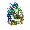
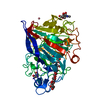
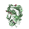
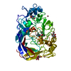
Entry Database : PDB / ID : 6grnTitle CELLOBIOHYDROLASE I (CEL7A) FROM Trichoderma reesei with S-dihydroxypropranolol in the active site Exoglucanase 1 Keywords / / / / Function / homology Function Domain/homology Component
/ / / / / / / / / / / / / / / / / / / / / / / / / / Biological species Trichoderma reesei (fungus)Method / / / Resolution : 1.79 Å Authors Sandgren, M. / Fagerstrom, A. / Widmalm, G. / Stahlberg, J. Funding support Organization Grant number Country Swedish Research Council Knut and Alice Wallenberg Foundation
Journal : Chemistry / Year : 2018Title : Enantioselective Binding of Propranolol and Analogues Thereof to Cellobiohydrolase Cel7A.Authors : Hamark, C. / Pendrill, R. / Landstrom, J. / Dotson Fagerstrom, A. / Sandgren, M. / Stahlberg, J. / Widmalm, G. History Deposition Jun 11, 2018 Deposition site / Processing site Revision 1.0 Oct 10, 2018 Provider / Type Revision 1.1 Dec 26, 2018 Group / Database references / Category / citation_authorItem _citation.journal_volume / _citation.page_first ... _citation.journal_volume / _citation.page_first / _citation.page_last / _citation.title / _citation_author.identifier_ORCID Revision 1.2 Nov 20, 2019 Group / Category / struct_connRevision 2.0 Mar 11, 2020 Group / Polymer sequence / Category / entity_polyItem / _entity_poly.pdbx_seq_one_letter_code_canRevision 2.1 Jul 29, 2020 Group / Derived calculations / Structure summaryCategory chem_comp / entity ... chem_comp / entity / pdbx_chem_comp_identifier / pdbx_entity_nonpoly / pdbx_struct_conn_angle / struct_conn / struct_site / struct_site_gen Item _chem_comp.name / _entity.pdbx_description ... _chem_comp.name / _entity.pdbx_description / _pdbx_entity_nonpoly.name / _pdbx_struct_conn_angle.ptnr1_auth_seq_id / _pdbx_struct_conn_angle.ptnr1_label_atom_id / _pdbx_struct_conn_angle.ptnr1_label_seq_id / _pdbx_struct_conn_angle.ptnr3_auth_seq_id / _pdbx_struct_conn_angle.ptnr3_label_atom_id / _pdbx_struct_conn_angle.ptnr3_label_seq_id / _pdbx_struct_conn_angle.value / _struct_conn.conn_type_id / _struct_conn.id / _struct_conn.pdbx_dist_value / _struct_conn.pdbx_leaving_atom_flag / _struct_conn.pdbx_role / _struct_conn.ptnr1_auth_comp_id / _struct_conn.ptnr1_auth_seq_id / _struct_conn.ptnr1_label_asym_id / _struct_conn.ptnr1_label_atom_id / _struct_conn.ptnr1_label_comp_id / _struct_conn.ptnr1_label_seq_id / _struct_conn.ptnr2_auth_comp_id / _struct_conn.ptnr2_auth_seq_id / _struct_conn.ptnr2_label_asym_id / _struct_conn.ptnr2_label_atom_id / _struct_conn.ptnr2_label_comp_id / _struct_conn.ptnr2_symmetry Description / Provider / Type Revision 2.2 Jan 17, 2024 Group Data collection / Database references ... Data collection / Database references / Refinement description / Structure summary Category chem_comp / chem_comp_atom ... chem_comp / chem_comp_atom / chem_comp_bond / database_2 / pdbx_initial_refinement_model Item / _database_2.pdbx_DOI / _database_2.pdbx_database_accessionRevision 2.3 Oct 16, 2024 Group / Category / pdbx_modification_feature
Show all Show less
 Movie
Movie Controller

 Yorodumi
 Open data
 Basic information
 Components
 Keywords
 Function and homology information
 Trichoderma reesei (fungus)
 X-RAY DIFFRACTION /
  Authors
 Sweden, 2items
  Citation
 Structure visualization
 Downloads & links
 Other downloads
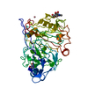


 PDBj
PDBj
 Assembly
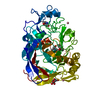

 Type: D-saccharide, beta linking / Mass: 221.208 Da / Num. of mol.: 1
 Mass: 58.933 Da / Num. of mol.: 2 / Source method: obtained synthetically / Formula: Co
 Mass: 291.342 Da / Num. of mol.: 1 / Source method: obtained synthetically / Formula: C16H21NO4 / Feature type: SUBJECT OF INVESTIGATION
 Mass: 18.015 Da / Num. of mol.: 470 / Source method: isolated from a natural source / Formula: H2O
 Sample preparation
 Processing