 Movie
Movie Controller

[English] 日本語
 Yorodumi
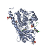
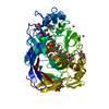
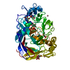
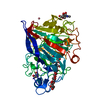
- PDB-1egn: CELLOBIOHYDROLASE CEL7A (E223S, A224H, L225V, T226A, D262G) MUTANT -
+ Open data

- Basic information
| Entry | Database: PDB / ID: 1egn | |||||||||
|---|---|---|---|---|---|---|---|---|---|---|
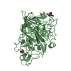
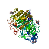
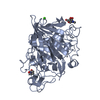
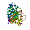
| Title | CELLOBIOHYDROLASE CEL7A (E223S, A224H, L225V, T226A, D262G) MUTANT | |||||||||
 Components Components | 1,4-BETA-D-GLUCAN CELLOBIOHYDROLASE CEL7A | |||||||||
 Keywords Keywords | HYDROLASE / GLYCOSIDASE / CELLULASE / CELLULOSE DEGRADATION / GLYCOPROTEIN / GLYCOSYLATED PROTEIN / PH-MUTANT | |||||||||
| Function / homology |  Function and homology information Function and homology informationcellulose 1,4-beta-cellobiosidase (non-reducing end) / cellulose 1,4-beta-cellobiosidase activity / cellulose binding / cellulose catabolic process / extracellular region Similarity search - Function | |||||||||
| Biological species |  Hypocrea jecorina (fungus) Hypocrea jecorina (fungus) | |||||||||
| Method |  X-RAY DIFFRACTION / SYNCHROTRON / Resolution: 1.6 Å X-RAY DIFFRACTION / SYNCHROTRON / Resolution: 1.6 Å | |||||||||
 Authors Authors | Stahlberg, J. / Harris, M. / Jones, T.A. | |||||||||
 Citation Citation | Journal: Biochem.J. / Year: 2001 Title: Engineering of a glycosidase Family 7 cellobiohydrolase to more alkaline pH optimum: the pH behaviour of Trichoderma reesei Cel7A and its E223S/ A224H/L225V/T226A/D262G mutant. Authors: Becker, D. / Braet, C. / Brumer III, H. / Claeyssens, M. / Divne, C. / Fagerstrom, B.R. / Harris, M. / Jones, T.A. / Kleywegt, G.J. / Koivula, A. / Mahdi, S. / Piens, K. / Sinnott, M.L. / ...Authors: Becker, D. / Braet, C. / Brumer III, H. / Claeyssens, M. / Divne, C. / Fagerstrom, B.R. / Harris, M. / Jones, T.A. / Kleywegt, G.J. / Koivula, A. / Mahdi, S. / Piens, K. / Sinnott, M.L. / Stahlberg, J. / Teeri, T.T. / Underwood, M. / Wohlfahrt, G. #1: Journal: Science / Year: 1994Title: The Three-dimensional Crystal Structure of the Catalytic Core of Cellobiohydrolase I from Trichoderma reesei Authors: Divne, C. / Stahlberg, J. / Reinikainen, T. / Ruohonen, L. / Pettersson, G. / Knowles, J.K. / Teeri, T.T. / Jones, T.A. #2: Journal: J.Mol.Biol. / Year: 1996Title: Activity Studies and Crystal Structures of Catalytically Deficient Mutants of Cellobiohydrolase I from Trichoderma reesei Authors: Stahlberg, J. / Divne, C. / Koivula, A. / Piens, K. / Claeyssens, M. / Teeri, T.T. / Jones, T.A. #3: Journal: J.Mol.Biol. / Year: 1998Title: High-resolution Crystal Structures Reveal how a Cellulose Chain is Bound in the 50a Long Tunnel of Cellobiohydrolase i from Trichoderma reesei Authors: Divne, C. / Stahlberg, J. / Teeri, T.T. / Jones, T.A. | |||||||||
| History |
|
- Structure visualization
| Structure viewer | Molecule: MolmilJmol/JSmol |
|---|
- Downloads & links
-Download
| PDBx/mmCIF format | 1egn.cif.gz | 101.3 KB | Display | PDBx/mmCIF format |
|---|---|---|---|---|
| PDB format | pdb1egn.ent.gz | 76.5 KB | Display | PDB format |
| PDBx/mmJSON format | 1egn.json.gz | Tree view | PDBx/mmJSON format | |
| Others |  Other downloads Other downloads |
-Validation report
| Arichive directory | https://data.pdbj.org/pub/pdb/validation_reports/eg/1egnftp://data.pdbj.org/pub/pdb/validation_reports/eg/1egn | HTTPS FTP |
|---|
-Related structure data


-Links
 PDBj
PDBj
- Assembly
| Deposited unit | 
| ||||||||||||
|---|---|---|---|---|---|---|---|---|---|---|---|---|---|
| 1 |
| ||||||||||||
| Unit cell |
| ||||||||||||
| Components on special symmetry positions |
|
-Components
| #1: Protein | Mass: 45991.680 Da / Num. of mol.: 1 / Fragment: CATALYTIC DOMAIN, RESIDUES 1 - 434 / Mutation: E223S, A224H, L225V, T226A, D262G Source method: isolated from a genetically manipulated source Source: (gene. exp.) Hypocrea jecorina (fungus) / Plasmid: PEM-F5 / Production host: Hypocrea jecorina (fungus)References: UniProt: P62694, cellulose 1,4-beta-cellobiosidase (non-reducing end) | ||||
|---|---|---|---|---|---|
| #2: Sugar | ChemComp-NAG /   Type: D-saccharide, beta linking / Mass: 221.208 Da / Num. of mol.: 1 Type: D-saccharide, beta linking / Mass: 221.208 Da / Num. of mol.: 1Source method: isolated from a genetically manipulated source Formula: C8H15NO6 | ||||
| #3: Chemical |   Mass: 58.933 Da / Num. of mol.: 2 / Source method: obtained synthetically / Formula: Co Mass: 58.933 Da / Num. of mol.: 2 / Source method: obtained synthetically / Formula: Co#4: Water | ChemComp-HOH / |  Mass: 18.015 Da / Num. of mol.: 354 / Source method: isolated from a natural source / Formula: H2O Mass: 18.015 Da / Num. of mol.: 354 / Source method: isolated from a natural source / Formula: H2OHas protein modification | Y | |
-Experimental details
-Experiment
| Experiment | Method: X-RAY DIFFRACTION / Number of used crystals: 1 |
|---|
- Sample preparation
| Crystal | Density Matthews: 2.08 Å3/Da / Density % sol: 40.9 % | ||||||||||||||||||||||||||||||||||||||||||
|---|---|---|---|---|---|---|---|---|---|---|---|---|---|---|---|---|---|---|---|---|---|---|---|---|---|---|---|---|---|---|---|---|---|---|---|---|---|---|---|---|---|---|---|
| Crystal grow | Temperature: 293 K / Method: vapor diffusion, hanging drop / pH: 5 Details: sodium acetate, PEG 5000 monomethyl-ether, glycerol, sodium morpholine-ethane-sulphonic acid, cobalt chloride , pH 5.0, VAPOR DIFFUSION, HANGING DROP, temperature 293K | ||||||||||||||||||||||||||||||||||||||||||
| Crystal grow | *PLUS | ||||||||||||||||||||||||||||||||||||||||||
| Components of the solutions | *PLUS
|
-Data collection
| Diffraction | Mean temperature: 100 K |
|---|---|
| Diffraction source | Source: SYNCHROTRON / Site: EMBL/DESY, HAMBURG  / Beamline: X11 / Wavelength: 0.905 / Beamline: X11 / Wavelength: 0.905 |
| Detector | Type: MARRESEARCH / Detector: IMAGE PLATE / Date: May 30, 1998 |
| Radiation | Protocol: SINGLE WAVELENGTH / Monochromatic (M) / Laue (L): M / Scattering type: x-ray |
| Radiation wavelength | Wavelength: 0.905 Å / Relative weight: 1 |
| Reflection | Resolution: 1.6→20 Å / Num. all: 50885 / Num. obs: 50885 / % possible obs: 99.8 % / Observed criterion σ(F): 0 / Observed criterion σ(I): 0 / Redundancy: 4.7 % / Biso Wilson estimate: 13.3 Å2 / Rmerge(I) obs: 0.064 / Net I/σ(I): 24.6 |
| Reflection shell | Resolution: 1.6→1.63 Å / Redundancy: 4.5 % / Rmerge(I) obs: 0.237 / % possible all: 99.9 |
| Reflection | *PLUS Lowest resolution: 30 Å / Num. obs: 50924 / % possible obs: 100 % |
| Reflection shell | *PLUS % possible obs: 99.9 % / Mean I/σ(I) obs: 5.5 |
- Processing
| Software |
| |||||||||||||||||||||||||
|---|---|---|---|---|---|---|---|---|---|---|---|---|---|---|---|---|---|---|---|---|---|---|---|---|---|---|
| Refinement | Resolution: 1.6→20 Å / σ(F): 0 / σ(I): 0 / Stereochemistry target values: Engh & Huber
| |||||||||||||||||||||||||
| Refinement step | Cycle: LAST / Resolution: 1.6→20 Å
| |||||||||||||||||||||||||
| Refine LS restraints |
| |||||||||||||||||||||||||
| Software | *PLUS Name: CNS / Classification: refinement | |||||||||||||||||||||||||
| Refinement | *PLUS Highest resolution: 1.6 Å / Lowest resolution: 20 Å / σ(F): 0 / Rfactor obs: 0.24 / Rfactor Rfree: 0.27 / Rfactor Rwork: 0.24 | |||||||||||||||||||||||||
| Solvent computation | *PLUS | |||||||||||||||||||||||||
| Displacement parameters | *PLUS | |||||||||||||||||||||||||
| Refine LS restraints | *PLUS
|
