 Movie
Movie Controller


+ Open data

- Basic information
| Entry | Database: PDB / ID: 5cel | ||||||||||||
|---|---|---|---|---|---|---|---|---|---|---|---|---|---|
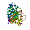
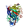
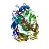
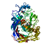
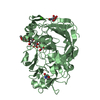
| Title | CBH1 (E212Q) CELLOTETRAOSE COMPLEX | ||||||||||||
 Components Components | 1,4-BETA-D-GLUCAN CELLOBIOHYDROLASE I | ||||||||||||
 Keywords Keywords | HYDROLASE / CELLULOSE DEGRADATION / GLYCOSIDASE / GLYCOPROTEIN / GLYCOSYLATED PROTEIN | ||||||||||||
| Function / homology |  Function and homology information Function and homology informationcellulose 1,4-beta-cellobiosidase (non-reducing end) / cellulose 1,4-beta-cellobiosidase activity / cellulose binding / cellulose catabolic process / extracellular region Similarity search - Function | ||||||||||||
| Biological species |  Hypocrea jecorina (fungus) Hypocrea jecorina (fungus) | ||||||||||||
| Method |  X-RAY DIFFRACTION / SYNCHROTRON / Resolution: 1.9 Å X-RAY DIFFRACTION / SYNCHROTRON / Resolution: 1.9 Å | ||||||||||||
 Authors Authors | Divne, C. / Stahlberg, J. / Jones, T.A. | ||||||||||||
 Citation Citation | Journal: J.Mol.Biol. / Year: 1998 Title: High-resolution crystal structures reveal how a cellulose chain is bound in the 50 A long tunnel of cellobiohydrolase I from Trichoderma reesei. Authors: Divne, C. / Stahlberg, J. / Teeri, T.T. / Jones, T.A. #1: Journal: J.Mol.Biol. / Year: 1996Title: Activity Studies and Crystal Structures of Catalytically Deficient Mutants of Cellobiohydrolase I from Trichoderma Reesei Authors: Stahlberg, J. / Divne, C. / Koivula, A. / Piens, K. / Claeyssens, M. / Teeri, T.T. / Jones, T.A. #2: Journal: Science / Year: 1994Title: The Three-Dimensional Crystal Structure of the Catalytic Core of Cellobiohydrolase I from Trichoderma Reesei Authors: Divne, C. / Stahlberg, J. / Reinikainen, T. / Ruohonen, L. / Pettersson, G. / Knowles, J.K. / Teeri, T.T. / Jones, T.A. #3: Journal: J.Mol.Biol. / Year: 1993Title: Crystallization and Preliminary X-Ray Studies on the Core Proteins of Cellobiohydrolase I and Endoglucanase I from Trichoderma Reesei Authors: Divne, C. / Sinning, I. / Stahlberg, J. / Pettersson, G. / Bailey, M. / Siika-Aho, M. / Margolles-Clark, E. / Teeri, T. / Jones, T.A. | ||||||||||||
| History |
|
- Structure visualization
| Structure viewer | Molecule: MolmilJmol/JSmol |
|---|
- Downloads & links
-Download
| PDBx/mmCIF format | 5cel.cif.gz | 102.5 KB | Display | PDBx/mmCIF format |
|---|---|---|---|---|
| PDB format | pdb5cel.ent.gz | 76.6 KB | Display | PDB format |
| PDBx/mmJSON format | 5cel.json.gz | Tree view | PDBx/mmJSON format | |
| Others |  Other downloads Other downloads |
-Validation report
| Arichive directory | https://data.pdbj.org/pub/pdb/validation_reports/ce/5celftp://data.pdbj.org/pub/pdb/validation_reports/ce/5cel | HTTPS FTP |
|---|
-Related structure data


-Links
 PDBj
PDBj
- Assembly
| Deposited unit | 
| ||||||||
|---|---|---|---|---|---|---|---|---|---|
| 1 |
| ||||||||
| Unit cell |
| ||||||||
| Components on special symmetry positions |
|
-Components
| #1: Protein | Mass: 46067.754 Da / Num. of mol.: 1 / Fragment: CATALYTIC DOMAIN, RESIDUES 1 - 434 / Mutation: E212Q Source method: isolated from a genetically manipulated source Source: (gene. exp.) Hypocrea jecorina (fungus) / Strain: QM 9414 / Gene: CBH1 / Variant: VTT-D-93201 / Plasmid: PEM-F5 / Gene (production host): CBH1 / Production host: Hypocrea jecorina (fungus)References: UniProt: P00725, UniProt: P62694*PLUS, cellulose 1,4-beta-cellobiosidase (non-reducing end) | ||||||||||||
|---|---|---|---|---|---|---|---|---|---|---|---|---|---|
| #2: Polysaccharide | 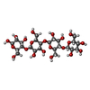  Source method: isolated from a genetically manipulated source Details: oligosaccharide / References: beta-cellotetraose #3: Sugar | ChemComp-NAG / |   Type: D-saccharide, beta linking / Mass: 221.208 Da / Num. of mol.: 1 Type: D-saccharide, beta linking / Mass: 221.208 Da / Num. of mol.: 1Source method: isolated from a genetically manipulated source Formula: C8H15NO6 #4: Chemical |   Mass: 58.933 Da / Num. of mol.: 2 / Source method: obtained synthetically / Formula: Co Mass: 58.933 Da / Num. of mol.: 2 / Source method: obtained synthetically / Formula: Co#5: Water | ChemComp-HOH / |  Mass: 18.015 Da / Num. of mol.: 431 / Source method: isolated from a natural source / Formula: H2O Mass: 18.015 Da / Num. of mol.: 431 / Source method: isolated from a natural source / Formula: H2OCompound details | THE COORDINATES GIVEN DEFINE THE STRUCTURE OF ONLY THE CATALYTIC DOMAIN (RESIDUES 1 - 434) OF THE ...THE COORDINATE | Has protein modification | Y | Nonpolymer details | THERE ARE TWO CELLOTETRAOSE MOLECULES BOUND IN THE CELLULOSE BINDING TUNNEL OF CBH1. THE ...THERE ARE TWO CELLOTETRA | |
-Experimental details
-Experiment
| Experiment | Method: X-RAY DIFFRACTION / Number of used crystals: 1 |
|---|
- Sample preparation
| Crystal | Density Matthews: 2.1 Å3/Da / Density % sol: 41 % | ||||||||||||||||||||||||||||||||||||||||||
|---|---|---|---|---|---|---|---|---|---|---|---|---|---|---|---|---|---|---|---|---|---|---|---|---|---|---|---|---|---|---|---|---|---|---|---|---|---|---|---|---|---|---|---|
| Crystal grow | Method: vapor diffusion, hanging drop / pH: 6 Details: HANGING DROPS. EQUAL VOLUMES OF 8 MG/ML PROTEIN AND RESERVOIR SOLUTION CONTAINING 0.1 M MES (PH 6.0), 18% (W/V) MONOMETHYLETHER PEG 5000, 0.01 M COCL2, AND 0.02% NA-AZIDE. ...Details: HANGING DROPS. EQUAL VOLUMES OF 8 MG/ML PROTEIN AND RESERVOIR SOLUTION CONTAINING 0.1 M MES (PH 6.0), 18% (W/V) MONOMETHYLETHER PEG 5000, 0.01 M COCL2, AND 0.02% NA-AZIDE. CRYOPROTECTANT/SOAK SOLUTION CONTAINED 0.1 M MES (PH 6.0), 20% (W/V) MONOMETHYLETHER PEG 5000, 0.01 M COCL2, 20% GLYCEROL AND 0.005 M CELLOTETRAOSE. THE AXES OF THE CRYO-COOLED CRYSTALS ARE SYSTEMATICALLY SHORTER THAN THOSE OF CRYSTALS COLLECTED AT ROOM TEMPERATURE. IN ORDER TO KEEP THE SAME INDEXING AS IN PREVIOUS ROOM-TEMPERATURE DATA SETS, THE LONGER A-AXIS IS LISTED BEFORE THE SHORTER B-AXIS., vapor diffusion - hanging drop | ||||||||||||||||||||||||||||||||||||||||||
| Crystal grow | *PLUS Method: vapor diffusion, hanging dropDetails: drop solution was mixed with an equal volume of reservoir solution | ||||||||||||||||||||||||||||||||||||||||||
| Components of the solutions | *PLUS
|
-Data collection
| Diffraction | Mean temperature: 120 K |
|---|---|
| Diffraction source | Source: SYNCHROTRON / Site: EMBL/DESY, HAMBURG  / Beamline: BW7B / Wavelength: 0.862 / Beamline: BW7B / Wavelength: 0.862 |
| Detector | Type: MARRESEARCH / Detector: IMAGE PLATE / Date: Sep 2, 1995 |
| Radiation | Monochromatic (M) / Laue (L): M / Scattering type: x-ray |
| Radiation wavelength | Wavelength: 0.862 Å / Relative weight: 1 |
| Reflection | Resolution: 1.9→65.9 Å / Num. obs: 29299 / % possible obs: 96.4 % / Observed criterion σ(I): 0 / Redundancy: 4.6 % / Rmerge(I) obs: 0.067 / Net I/σ(I): 5 |
| Reflection shell | Resolution: 1.9→1.95 Å / Redundancy: 3.6 % / Rmerge(I) obs: 0.402 / Mean I/σ(I) obs: 1.5 / % possible all: 92.7 |
| Reflection shell | *PLUS % possible obs: 92.7 % |
- Processing
| Software |
| ||||||||||||||||||||||||||||||||||||||||||||||||||||||||||||
|---|---|---|---|---|---|---|---|---|---|---|---|---|---|---|---|---|---|---|---|---|---|---|---|---|---|---|---|---|---|---|---|---|---|---|---|---|---|---|---|---|---|---|---|---|---|---|---|---|---|---|---|---|---|---|---|---|---|---|---|---|---|
| Refinement | Resolution: 1.9→7.5 Å / σ(F): 0
| ||||||||||||||||||||||||||||||||||||||||||||||||||||||||||||
| Displacement parameters | Biso mean: 7.9 Å2 | ||||||||||||||||||||||||||||||||||||||||||||||||||||||||||||
| Refinement step | Cycle: LAST / Resolution: 1.9→7.5 Å
| ||||||||||||||||||||||||||||||||||||||||||||||||||||||||||||
| Refine LS restraints |
| ||||||||||||||||||||||||||||||||||||||||||||||||||||||||||||
| Xplor file |
| ||||||||||||||||||||||||||||||||||||||||||||||||||||||||||||
| Software | *PLUS Name: X-PLOR / Version: 3.1 / Classification: refinement | ||||||||||||||||||||||||||||||||||||||||||||||||||||||||||||
| Refinement | *PLUS | ||||||||||||||||||||||||||||||||||||||||||||||||||||||||||||
| Solvent computation | *PLUS | ||||||||||||||||||||||||||||||||||||||||||||||||||||||||||||
| Displacement parameters | *PLUS |
