 Movie
Movie Controller


+ Open data

- Basic information
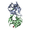
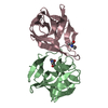
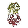
| Entry | Database: PDB / ID: 6gkq | ||||||
|---|---|---|---|---|---|---|---|
| Title | X-ray structure determined from ex vivo Charcot-Leyden crystal | ||||||
 Components Components | Galectin-10 | ||||||
 Keywords Keywords | SUGAR BINDING PROTEIN / Galectin-10 / ex vivo crystal | ||||||
| Function / homology |  Function and homology information Function and homology informationregulation of activated T cell proliferation / regulation of T cell cytokine production / T cell apoptotic process / regulation of T cell anergy / carbohydrate binding / extracellular matrix / identical protein binding / cytosol Similarity search - Function | ||||||
| Biological species |  Homo sapiens (human) Homo sapiens (human) | ||||||
| Method |  X-RAY DIFFRACTION / SYNCHROTRON / MOLECULAR REPLACEMENT / Resolution: 2.3 Å X-RAY DIFFRACTION / SYNCHROTRON / MOLECULAR REPLACEMENT / Resolution: 2.3 Å | ||||||
 Authors Authors | Verstraete, K. / Verschueren, K. / Savvides, S.N. | ||||||
 Citation Citation | Journal: Science / Year: 2019 Title: Protein crystallization promotes type 2 immunity and is reversible by antibody treatment. Authors: Emma K Persson / Kenneth Verstraete / Ines Heyndrickx / Elien Gevaert / Helena Aegerter / Jean-Michel Percier / Kim Deswarte / Koen H G Verschueren / Ann Dansercoer / Delphine Gras / Pascal ...Authors: Emma K Persson / Kenneth Verstraete / Ines Heyndrickx / Elien Gevaert / Helena Aegerter / Jean-Michel Percier / Kim Deswarte / Koen H G Verschueren / Ann Dansercoer / Delphine Gras / Pascal Chanez / Claus Bachert / Amanda Gonçalves / Hanne Van Gorp / Hans De Haard / Christophe Blanchetot / Michael Saunders / Hamida Hammad / Savvas N Savvides / Bart N Lambrecht /     Abstract: Although spontaneous protein crystallization is a rare event in vivo, Charcot-Leyden crystals (CLCs) consisting of galectin-10 (Gal10) protein are frequently observed in eosinophilic diseases, such ...Although spontaneous protein crystallization is a rare event in vivo, Charcot-Leyden crystals (CLCs) consisting of galectin-10 (Gal10) protein are frequently observed in eosinophilic diseases, such as asthma. We found that CLCs derived from patients showed crystal packing and Gal10 structure identical to those of Gal10 crystals grown in vitro. When administered to the airways, crystalline Gal10 stimulated innate and adaptive immunity and acted as a type 2 adjuvant. By contrast, a soluble Gal10 mutein was inert. Antibodies directed against key epitopes of the CLC crystallization interface dissolved preexisting CLCs in patient-derived mucus within hours and reversed crystal-driven inflammation, goblet-cell metaplasia, immunoglobulin E (IgE) synthesis, and bronchial hyperreactivity (BHR) in a humanized mouse model of asthma. Thus, protein crystals may promote hallmark features of asthma and are targetable by crystal-dissolving antibodies. | ||||||
| History |
|
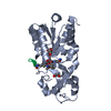
- Structure visualization
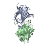
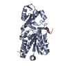
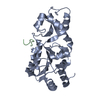
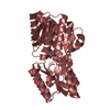
| Structure viewer | Molecule: MolmilJmol/JSmol |
|---|
- Downloads & links
-Download
| PDBx/mmCIF format | 6gkq.cif.gz | 73.4 KB | Display | PDBx/mmCIF format |
|---|---|---|---|---|
| PDB format | pdb6gkq.ent.gz | 54.2 KB | Display | PDB format |
| PDBx/mmJSON format | 6gkq.json.gz | Tree view | PDBx/mmJSON format | |
| Others |  Other downloads Other downloads |
-Validation report
| Arichive directory | https://data.pdbj.org/pub/pdb/validation_reports/gk/6gkqftp://data.pdbj.org/pub/pdb/validation_reports/gk/6gkq | HTTPS FTP |
|---|
-Related structure data
| Related structure data |  6gksC  6gktC 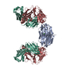 6gkuC  6glwC 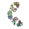 6glxC 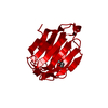 6qrnC  1lclS S: Starting model for refinement C: citing same article ( |
|---|---|
| Similar structure data |


-Links
 PDBj
PDBj- Assembly
| Deposited unit | 
| |||||||||
|---|---|---|---|---|---|---|---|---|---|---|
| 1 | 
| |||||||||
| Unit cell |
| |||||||||
| Components on special symmetry positions |
|
-Components
| #1: Protein | Mass: 16499.887 Da / Num. of mol.: 1 / Mutation: Ala28Val (Natural variant) / Source method: isolated from a natural source Details: This X-ray data was collected from an ex vivo Charcot-Leyden crystal. Source: (natural) Homo sapiens (human) / References: UniProt: Q05315 |
|---|---|
| #2: Chemical | ChemComp-GOL /   Mass: 92.094 Da / Num. of mol.: 1 / Source method: obtained synthetically / Formula: C3H8O3 Mass: 92.094 Da / Num. of mol.: 1 / Source method: obtained synthetically / Formula: C3H8O3 |
| #3: Water | ChemComp-HOH /  Mass: 18.015 Da / Num. of mol.: 112 / Source method: isolated from a natural source / Formula: H2O Mass: 18.015 Da / Num. of mol.: 112 / Source method: isolated from a natural source / Formula: H2O |
-Experimental details
-Experiment
| Experiment | Method: X-RAY DIFFRACTION / Number of used crystals: 1 |
|---|
- Sample preparation
| Crystal | Density Matthews: 2.71 Å3/Da / Density % sol: 54.55 % |
|---|---|
| Crystal grow | Temperature: 310 K / Method: in cell Details: Data was collected from ex vivo Charcot-Leyden crystals. Sticky allergic mucin type mucus was obtained from CRSwNP patients undergoing endoscopic sinus surgery. One g of the allergic mucin ...Details: Data was collected from ex vivo Charcot-Leyden crystals. Sticky allergic mucin type mucus was obtained from CRSwNP patients undergoing endoscopic sinus surgery. One g of the allergic mucin was cut thoroughly in 10 ml RPMI-1640 (Sigma-Aldrich) containing antibiotics (50 IU/mL penicillin and 50 mg/mL streptomycin; One g of the allergic mucin was cut thoroughly in 10 ml RPMI-1640 (Sigma-Aldrich) containing antibiotics (50 IU/mL penicillin and 50 mg/mL streptomycin; Thermo Fisher Scientific), 0.1 percent BSA (Sigma-Aldrich) and 1 mg/ml Collagen type 2 (Worthington), and further homogenized using a GentleMACS Dissociator (Myltenyi Biotec) and subsequently incubed at 37 degrees for 45 minutes under continuous rotation. After centrifugation the pellet was dissolved in 3 ml PBS containing antibiotics, to which 6 ml Ficoll-Paque (GE Healthcare) was added. After centrifugation at 250g and removal of the supernatant and most of the Ficoll layer, the pellet was dissolved 1:10 in PBS with antibiotics. This precipitation process was repeated 5 more times. The final pellet containing the crystals was resuspended in PBS with antibiotics. |
-Data collection
| Diffraction | Mean temperature: 100 K |
|---|---|
| Diffraction source | Source: SYNCHROTRON / Site: PETRA III, EMBL c/o DESY  / Beamline: P14 (MX2) / Wavelength: 0.9763 Å / Beamline: P14 (MX2) / Wavelength: 0.9763 Å |
| Detector | Type: DECTRIS EIGER X 16M / Detector: PIXEL / Date: Sep 22, 2015 |
| Radiation | Protocol: SINGLE WAVELENGTH / Monochromatic (M) / Laue (L): M / Scattering type: x-ray |
| Radiation wavelength | Wavelength: 0.9763 Å / Relative weight: 1 |
| Reflection | Resolution: 2.22→50 Å / Num. obs: 9972 / % possible obs: 99.9 % / Redundancy: 11.86 % / Biso Wilson estimate: 23.36 Å2 / CC1/2: 0.99 / Rrim(I) all: 0.31 / Net I/σ(I): 7.85 |
| Reflection shell | Resolution: 2.22→2.35 Å / Redundancy: 12.2 % / Mean I/σ(I) obs: 1.29 / Num. unique obs: 1533 / CC1/2: 0.48 / Rrim(I) all: 0.195 / % possible all: 99.1 |
- Processing
| Software |
| ||||||||||||||||||||||||||||||||||||||||||||||||||||||||||||||||||||||||||||||||||||||||||||||||||||||||||||||||||||||||||||||||||||||||||||||||||||||||||||||||||||||||||||||||||||||||||||||||||||||||
|---|---|---|---|---|---|---|---|---|---|---|---|---|---|---|---|---|---|---|---|---|---|---|---|---|---|---|---|---|---|---|---|---|---|---|---|---|---|---|---|---|---|---|---|---|---|---|---|---|---|---|---|---|---|---|---|---|---|---|---|---|---|---|---|---|---|---|---|---|---|---|---|---|---|---|---|---|---|---|---|---|---|---|---|---|---|---|---|---|---|---|---|---|---|---|---|---|---|---|---|---|---|---|---|---|---|---|---|---|---|---|---|---|---|---|---|---|---|---|---|---|---|---|---|---|---|---|---|---|---|---|---|---|---|---|---|---|---|---|---|---|---|---|---|---|---|---|---|---|---|---|---|---|---|---|---|---|---|---|---|---|---|---|---|---|---|---|---|---|---|---|---|---|---|---|---|---|---|---|---|---|---|---|---|---|---|---|---|---|---|---|---|---|---|---|---|---|---|---|---|---|---|
| Refinement | Method to determine structure: MOLECULAR REPLACEMENT Starting model: 1LCL Resolution: 2.3→43.06 Å / Cor.coef. Fo:Fc: 0.919 / Cor.coef. Fo:Fc free: 0.878 / SU R Cruickshank DPI: 0.237 / Cross valid method: THROUGHOUT / σ(F): 0 / SU R Blow DPI: 0.304 / SU Rfree Blow DPI: 0.215 / SU Rfree Cruickshank DPI: 0.197
| ||||||||||||||||||||||||||||||||||||||||||||||||||||||||||||||||||||||||||||||||||||||||||||||||||||||||||||||||||||||||||||||||||||||||||||||||||||||||||||||||||||||||||||||||||||||||||||||||||||||||
| Displacement parameters | Biso mean: 29.85 Å2
| ||||||||||||||||||||||||||||||||||||||||||||||||||||||||||||||||||||||||||||||||||||||||||||||||||||||||||||||||||||||||||||||||||||||||||||||||||||||||||||||||||||||||||||||||||||||||||||||||||||||||
| Refine analyze | Luzzati coordinate error obs: 0.28 Å | ||||||||||||||||||||||||||||||||||||||||||||||||||||||||||||||||||||||||||||||||||||||||||||||||||||||||||||||||||||||||||||||||||||||||||||||||||||||||||||||||||||||||||||||||||||||||||||||||||||||||
| Refinement step | Cycle: 1 / Resolution: 2.3→43.06 Å
| ||||||||||||||||||||||||||||||||||||||||||||||||||||||||||||||||||||||||||||||||||||||||||||||||||||||||||||||||||||||||||||||||||||||||||||||||||||||||||||||||||||||||||||||||||||||||||||||||||||||||
| Refine LS restraints |
| ||||||||||||||||||||||||||||||||||||||||||||||||||||||||||||||||||||||||||||||||||||||||||||||||||||||||||||||||||||||||||||||||||||||||||||||||||||||||||||||||||||||||||||||||||||||||||||||||||||||||
| LS refinement shell | Resolution: 2.3→2.57 Å / Rfactor Rfree error: 0 / Total num. of bins used: 5
| ||||||||||||||||||||||||||||||||||||||||||||||||||||||||||||||||||||||||||||||||||||||||||||||||||||||||||||||||||||||||||||||||||||||||||||||||||||||||||||||||||||||||||||||||||||||||||||||||||||||||
| Refinement TLS params. | Method: refined / Refine-ID: X-RAY DIFFRACTION
| ||||||||||||||||||||||||||||||||||||||||||||||||||||||||||||||||||||||||||||||||||||||||||||||||||||||||||||||||||||||||||||||||||||||||||||||||||||||||||||||||||||||||||||||||||||||||||||||||||||||||
| Refinement TLS group |
|
