 Movie
Movie Controller


+ Open data

- Basic information
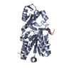
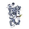
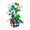
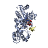
| Entry | Database: PDB / ID: 2h2f | ||||||
|---|---|---|---|---|---|---|---|
| Title | The Structural basis for Sirtuin Substrate affinity | ||||||
 Components Components |
| ||||||
 Keywords Keywords | HYDROLASE / Sir2Tm | ||||||
| Function / homology |  Function and homology information Function and homology informationprotein acetyllysine N-acetyltransferase / NAD-dependent protein lysine deacetylase activity / negative regulation of helicase activity / Loss of function of TP53 in cancer due to loss of tetramerization ability / Regulation of TP53 Expression / signal transduction by p53 class mediator / negative regulation of G1 to G0 transition / negative regulation of glucose catabolic process to lactate via pyruvate / Transcriptional activation of cell cycle inhibitor p21 / regulation of intrinsic apoptotic signaling pathway by p53 class mediator ...protein acetyllysine N-acetyltransferase / NAD-dependent protein lysine deacetylase activity / negative regulation of helicase activity / Loss of function of TP53 in cancer due to loss of tetramerization ability / Regulation of TP53 Expression / signal transduction by p53 class mediator / negative regulation of G1 to G0 transition / negative regulation of glucose catabolic process to lactate via pyruvate / Transcriptional activation of cell cycle inhibitor p21 / regulation of intrinsic apoptotic signaling pathway by p53 class mediator / negative regulation of pentose-phosphate shunt / Activation of NOXA and translocation to mitochondria / ATP-dependent DNA/DNA annealing activity / regulation of cell cycle G2/M phase transition / oligodendrocyte apoptotic process / negative regulation of miRNA processing / intrinsic apoptotic signaling pathway in response to hypoxia / oxidative stress-induced premature senescence / regulation of tissue remodeling / positive regulation of thymocyte apoptotic process / positive regulation of mitochondrial membrane permeability / germ cell nucleus / regulation of fibroblast apoptotic process / bone marrow development / cellular response to actinomycin D / circadian behavior / histone deacetylase regulator activity / positive regulation of programmed necrotic cell death / : / regulation of mitochondrial membrane permeability involved in apoptotic process / RUNX3 regulates CDKN1A transcription / T cell proliferation involved in immune response / TP53 Regulates Transcription of Death Receptors and Ligands / Activation of PUMA and translocation to mitochondria / TP53 regulates transcription of additional cell cycle genes whose exact role in the p53 pathway remain uncertain / mRNA transcription / negative regulation of glial cell proliferation / negative regulation of neuroblast proliferation / regulation of DNA damage response, signal transduction by p53 class mediator / Regulation of TP53 Activity through Association with Co-factors / Formation of Senescence-Associated Heterochromatin Foci (SAHF) / mitochondrial DNA repair / T cell lineage commitment / thymocyte apoptotic process / ER overload response / TP53 Regulates Transcription of Caspase Activators and Caspases / cardiac septum morphogenesis / B cell lineage commitment / entrainment of circadian clock by photoperiod / negative regulation of DNA replication / Zygotic genome activation (ZGA) / negative regulation of mitophagy / TP53 Regulates Transcription of Genes Involved in Cytochrome C Release / necroptotic process / PI5P Regulates TP53 Acetylation / Association of TriC/CCT with target proteins during biosynthesis / positive regulation of release of cytochrome c from mitochondria / negative regulation of telomere maintenance via telomerase / SUMOylation of transcription factors / TP53 regulates transcription of several additional cell death genes whose specific roles in p53-dependent apoptosis remain uncertain / rRNA transcription / negative regulation of reactive oxygen species metabolic process / intrinsic apoptotic signaling pathway by p53 class mediator / TFIID-class transcription factor complex binding / Transcriptional Regulation by VENTX / cellular response to UV-C / viral process / neuroblast proliferation / NAD+ poly-ADP-ribosyltransferase activity / NAD+ binding / intrinsic apoptotic signaling pathway in response to endoplasmic reticulum stress / replicative senescence / intrinsic apoptotic signaling pathway in response to DNA damage by p53 class mediator / Pyroptosis / positive regulation of RNA polymerase II transcription preinitiation complex assembly / chromosome organization / general transcription initiation factor binding / positive regulation of execution phase of apoptosis / hematopoietic stem cell differentiation / embryonic organ development / type II interferon-mediated signaling pathway / TP53 Regulates Transcription of Genes Involved in G1 Cell Cycle Arrest / response to X-ray / hematopoietic progenitor cell differentiation / somitogenesis / negative regulation of stem cell proliferation / core promoter sequence-specific DNA binding / glial cell proliferation / negative regulation of fibroblast proliferation / cis-regulatory region sequence-specific DNA binding / cellular response to glucose starvation / mitophagy / Regulation of TP53 Activity through Acetylation / negative regulation of proteolysis / mitotic G1 DNA damage checkpoint signaling / positive regulation of intrinsic apoptotic signaling pathway / response to salt stress / cardiac muscle cell apoptotic process / transcription repressor complex / 14-3-3 protein binding Similarity search - Function | ||||||
| Biological species |   Thermotoga maritima (bacteria) Thermotoga maritima (bacteria) | ||||||
| Method |  X-RAY DIFFRACTION / SYNCHROTRON / MOLECULAR REPLACEMENT / Resolution: 2.2 Å X-RAY DIFFRACTION / SYNCHROTRON / MOLECULAR REPLACEMENT / Resolution: 2.2 Å | ||||||
 Authors Authors | Cosgrove, M.S. / Wolberger, C. | ||||||
 Citation Citation | Journal: Biochemistry / Year: 2006 Title: The structural basis of sirtuin substrate affinity Authors: Cosgrove, M.S. / Bever, K. / Avalos, J.L. / Muhammad, S. / Zhang, X. / Wolberger, C. | ||||||
| History |
|
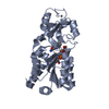
- Structure visualization
| Structure viewer | Molecule: MolmilJmol/JSmol |
|---|
- Downloads & links
-Download
| PDBx/mmCIF format | 2h2f.cif.gz | 62.4 KB | Display | PDBx/mmCIF format |
|---|---|---|---|---|
| PDB format | pdb2h2f.ent.gz | 44.6 KB | Display | PDB format |
| PDBx/mmJSON format | 2h2f.json.gz | Tree view | PDBx/mmJSON format | |
| Others |  Other downloads Other downloads |
-Validation report
| Arichive directory | https://data.pdbj.org/pub/pdb/validation_reports/h2/2h2fftp://data.pdbj.org/pub/pdb/validation_reports/h2/2h2f | HTTPS FTP |
|---|
-Related structure data
| Related structure data |  2h2dC 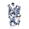 2h2gC 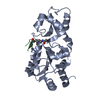 2h2hC  2h2iSC S: Starting model for refinement C: citing same article ( |
|---|---|
| Similar structure data |


-Links
 PDBj
PDBj


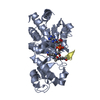
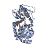
- Assembly
| Deposited unit | 
| ||||||||
|---|---|---|---|---|---|---|---|---|---|
| 1 |
| ||||||||
| Unit cell |
|
-Components
| #1: Protein | Mass: 27569.793 Da / Num. of mol.: 1 Source method: isolated from a genetically manipulated source Source: (gene. exp.) Thermotoga maritima (bacteria) / Gene: npdA / Production host: References: UniProt: Q9WYW0, Hydrolases; Acting on carbon-nitrogen bonds, other than peptide bonds; In linear amides |
|---|---|
| #2: Protein/peptide | Mass: 2098.491 Da / Num. of mol.: 1 / Source method: obtained synthetically Details: THE SEQUENCE OF THE PEPTIDE IS NATURALLY FOUND IN HOMO SAPIENS (HUMAN). References: UniProt: P04637 |
| #3: Chemical | ChemComp-ZN /   Mass: 65.409 Da / Num. of mol.: 1 / Source method: obtained synthetically / Formula: Zn Mass: 65.409 Da / Num. of mol.: 1 / Source method: obtained synthetically / Formula: Zn |
| #4: Water | ChemComp-HOH /  Mass: 18.015 Da / Num. of mol.: 97 / Source method: isolated from a natural source / Formula: H2O Mass: 18.015 Da / Num. of mol.: 97 / Source method: isolated from a natural source / Formula: H2O |
-Experimental details
-Experiment
| Experiment | Method: X-RAY DIFFRACTION / Number of used crystals: 1 |
|---|
- Sample preparation
| Crystal | Density Matthews: 2.42 Å3/Da / Density % sol: 49.09 % |
|---|---|
| Crystal grow | Temperature: 298 K / pH: 9.6 Details: 20% PEG, pH 9.6, VAPOR DIFFUSION, HANGING DROP, temperature 298K, pH 9.60 |
-Data collection
| Diffraction | Mean temperature: 100 K |
|---|---|
| Diffraction source | Source: SYNCHROTRON / Site: NSLS  / Beamline: X25 / Wavelength: 1.1 / Beamline: X25 / Wavelength: 1.1 |
| Detector | Detector: CCD / Date: Aug 14, 2004 |
| Radiation | Protocol: SINGLE WAVELENGTH / Monochromatic (M) / Laue (L): M / Scattering type: x-ray |
| Radiation wavelength | Wavelength: 1.1 Å / Relative weight: 1 |
| Reflection | Resolution: 2.1→50 Å / Num. obs: 17279 / % possible obs: 99.2 % / Observed criterion σ(I): 2 / Redundancy: 6.2 % / Biso Wilson estimate: 23.3 Å2 / Rmerge(I) obs: 0.092 / Rsym value: 0.065 / Net I/σ(I): 29.2 |
| Reflection shell | Resolution: 2.1→2.18 Å / Redundancy: 5.5 % / Rmerge(I) obs: 0.66 / Mean I/σ(I) obs: 3.7 / Rsym value: 0.559 / % possible all: 98.6 |
- Processing
| Software |
| ||||||||||||||||||||||||||||||||||||||||||||||||||||||||||||
|---|---|---|---|---|---|---|---|---|---|---|---|---|---|---|---|---|---|---|---|---|---|---|---|---|---|---|---|---|---|---|---|---|---|---|---|---|---|---|---|---|---|---|---|---|---|---|---|---|---|---|---|---|---|---|---|---|---|---|---|---|---|
| Refinement | Method to determine structure: MOLECULAR REPLACEMENT Starting model: PDB Entry: 2H2I Resolution: 2.2→7.96 Å / Rfactor Rfree error: 0.009 / Data cutoff high absF: 1289818.32 / Data cutoff low absF: 0 / Isotropic thermal model: RESTRAINED / Cross valid method: THROUGHOUT / σ(F): 0 / Stereochemistry target values: ENGH & HUBER
| ||||||||||||||||||||||||||||||||||||||||||||||||||||||||||||
| Solvent computation | Solvent model: FLAT / Bsol: 704696 Å2 / ksol: 0.579667 e/Å3 | ||||||||||||||||||||||||||||||||||||||||||||||||||||||||||||
| Displacement parameters | Biso mean: 38 Å2
| ||||||||||||||||||||||||||||||||||||||||||||||||||||||||||||
| Refine analyze |
| ||||||||||||||||||||||||||||||||||||||||||||||||||||||||||||
| Refinement step | Cycle: LAST / Resolution: 2.2→7.96 Å
| ||||||||||||||||||||||||||||||||||||||||||||||||||||||||||||
| Refine LS restraints |
| ||||||||||||||||||||||||||||||||||||||||||||||||||||||||||||
| LS refinement shell | Resolution: 2.2→2.33 Å / Rfactor Rfree error: 0.024 / Total num. of bins used: 6
|
