 Movie
Movie Controller


+ Open data

- Basic information
| Entry | Database: PDB / ID: 6d5n | |||||||||
|---|---|---|---|---|---|---|---|---|---|---|
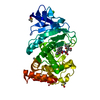
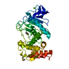
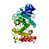
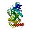
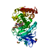
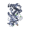
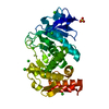
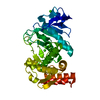
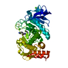
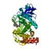
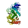
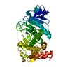
| Title | Hexagonal thermolysin (295) in the presence of 50% xylose | |||||||||
 Components Components | Thermolysin | |||||||||
 Keywords Keywords | HYDROLASE / zinc protease | |||||||||
| Function / homology |  Function and homology information Function and homology informationthermolysin / metalloendopeptidase activity / proteolysis / extracellular region / metal ion binding Similarity search - Function | |||||||||
| Biological species |  | |||||||||
| Method |  X-RAY DIFFRACTION / Resolution: 2.00003665634 Å X-RAY DIFFRACTION / Resolution: 2.00003665634 Å | |||||||||
 Authors Authors | Juers, D.H. | |||||||||
| Funding support |  United States, 1items United States, 1items
| |||||||||
 Citation Citation | Journal: Acta Crystallogr D Struct Biol / Year: 2018 Title: The impact of cryosolution thermal contraction on proteins and protein crystals: volumes, conformation and order. Authors: Juers, D.H. / Farley, C.A. / Saxby, C.P. / Cotter, R.A. / Cahn, J.K.B. / Holton-Burke, R.C. / Harrison, K. / Wu, Z. | |||||||||
| History |
|
- Structure visualization
| Structure viewer | Molecule: MolmilJmol/JSmol |
|---|
- Downloads & links
-Download
| PDBx/mmCIF format | 6d5n.cif.gz | 159.1 KB | Display | PDBx/mmCIF format |
|---|---|---|---|---|
| PDB format | pdb6d5n.ent.gz | 102.4 KB | Display | PDB format |
| PDBx/mmJSON format | 6d5n.json.gz | Tree view | PDBx/mmJSON format | |
| Others |  Other downloads Other downloads |
-Validation report
| Arichive directory | https://data.pdbj.org/pub/pdb/validation_reports/d5/6d5nftp://data.pdbj.org/pub/pdb/validation_reports/d5/6d5n | HTTPS FTP |
|---|
-Related structure data
| Related structure data | 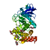 5un3C 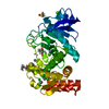 5uu7C 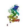 5uu8C  5uu9C 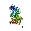 5uuaC 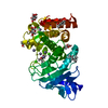 5uubC 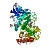 5uucC 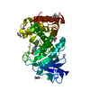 5uudC 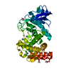 5uueC 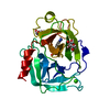 6avlC 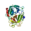 6b6nC 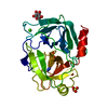 6b6oC 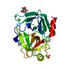 6b6pC 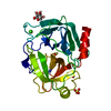 6b6qC 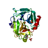 6b6rC  6b6sC 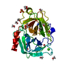 6b6tC 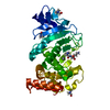 6d5oC  6d5pC  6d5qC  6d5rC  6d5sC 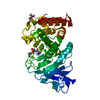 6d5tC 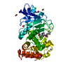 6d5uC 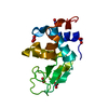 6d6eC 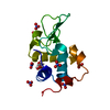 6d6fC 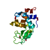 6d6gC 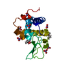 6d6hC 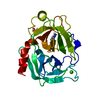 6dzfC C: citing same article ( |
|---|---|
| Similar structure data |


-Links
 PDBj
PDBj
- Assembly
| Deposited unit | 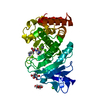
| ||||||||||||
|---|---|---|---|---|---|---|---|---|---|---|---|---|---|
| 1 |
| ||||||||||||
| Unit cell |
| ||||||||||||
| Components on special symmetry positions |
|
-Components
-Protein / Sugars , 2 types, 3 molecules A
| #1: Protein | Mass: 34360.336 Da / Num. of mol.: 1 / Source method: isolated from a natural source / Source: (natural) |
|---|---|
| #6: Sugar |  Type: D-saccharide, beta linking / Mass: 150.130 Da / Num. of mol.: 2 / Source method: obtained synthetically / Formula: C5H10O5 Type: D-saccharide, beta linking / Mass: 150.130 Da / Num. of mol.: 2 / Source method: obtained synthetically / Formula: C5H10O5 |
-Non-polymers , 5 types, 147 molecules 


| #2: Chemical | ChemComp-VAL /  Type: L-peptide linking / Mass: 117.146 Da / Num. of mol.: 1 / Source method: obtained synthetically / Formula: C5H11NO2 Type: L-peptide linking / Mass: 117.146 Da / Num. of mol.: 1 / Source method: obtained synthetically / Formula: C5H11NO2 | ||
|---|---|---|---|
| #3: Chemical | ChemComp-LYS /  Type: L-peptide linking / Mass: 147.195 Da / Num. of mol.: 1 / Source method: obtained synthetically / Formula: C6H15N2O2 Type: L-peptide linking / Mass: 147.195 Da / Num. of mol.: 1 / Source method: obtained synthetically / Formula: C6H15N2O2 | ||
| #4: Chemical | ChemComp-ZN /  Mass: 65.409 Da / Num. of mol.: 1 / Source method: obtained synthetically / Formula: Zn Mass: 65.409 Da / Num. of mol.: 1 / Source method: obtained synthetically / Formula: Zn | ||
| #5: Chemical | ChemComp-CA /  Mass: 40.078 Da / Num. of mol.: 4 / Source method: obtained synthetically / Formula: Ca Mass: 40.078 Da / Num. of mol.: 4 / Source method: obtained synthetically / Formula: Ca#7: Water | ChemComp-HOH / | Mass: 18.015 Da / Num. of mol.: 140 / Source method: isolated from a natural source / Formula: H2O |
-Experimental details
-Experiment
| Experiment | Method: X-RAY DIFFRACTION / Number of used crystals: 1 |
|---|
- Sample preparation
| Crystal | Density Matthews: 2.43 Å3/Da / Density % sol: 49.33 % |
|---|---|
| Crystal grow | Temperature: 295 K / Method: vapor diffusion, hanging drop / Details: Protein: 100 mg/mL in 45% DMSO Well: 2 M AmSO4 |
-Data collection
| Diffraction | Mean temperature: 295 K |
|---|---|
| Diffraction source | Source: ROTATING ANODE / Type: Agilent SuperNova / Wavelength: 1.54 Å |
| Detector | Type: OXFORD ONYX CCD / Detector: CCD / Date: Jul 19, 2016 |
| Radiation | Protocol: SINGLE WAVELENGTH / Monochromatic (M) / Laue (L): M / Scattering type: x-ray |
| Radiation wavelength | Wavelength: 1.54 Å / Relative weight: 1 |
| Reflection | Resolution: 2→13.86 Å / Num. obs: 23812 / % possible obs: 99.6 % / Redundancy: 5.7 % / Biso Wilson estimate: 19.6315777022 Å2 / CC1/2: 0.995 / Rrim(I) all: 0.142 / Net I/σ(I): 11.7 |
| Reflection shell | Resolution: 2→2.11 Å / CC1/2: 0.736 |
- Processing
| Software |
| |||||||||||||||||||||||||||||||||||||||||||||||||||||||||||||||
|---|---|---|---|---|---|---|---|---|---|---|---|---|---|---|---|---|---|---|---|---|---|---|---|---|---|---|---|---|---|---|---|---|---|---|---|---|---|---|---|---|---|---|---|---|---|---|---|---|---|---|---|---|---|---|---|---|---|---|---|---|---|---|---|---|
| Refinement | Resolution: 2.00003665634→13.8519097476 Å / SU ML: 0.213793487267 / Cross valid method: FREE R-VALUE / σ(F): 1.33721327711 / Phase error: 17.3279762667
| |||||||||||||||||||||||||||||||||||||||||||||||||||||||||||||||
| Solvent computation | Shrinkage radii: 0.9 Å / VDW probe radii: 1.11 Å | |||||||||||||||||||||||||||||||||||||||||||||||||||||||||||||||
| Displacement parameters | Biso mean: 22.9275337172 Å2 | |||||||||||||||||||||||||||||||||||||||||||||||||||||||||||||||
| Refinement step | Cycle: LAST / Resolution: 2.00003665634→13.8519097476 Å
| |||||||||||||||||||||||||||||||||||||||||||||||||||||||||||||||
| Refine LS restraints |
| |||||||||||||||||||||||||||||||||||||||||||||||||||||||||||||||
| LS refinement shell |
|
