 Movie
Movie Controller

[English] 日本語
 Yorodumi
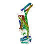
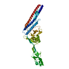
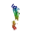
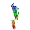
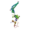
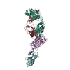
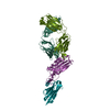
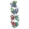
- PDB-6d32: Crystal structure of Xenopus Smoothened in complex with cyclopamine -
+ Open data

- Basic information
| Entry | Database: PDB / ID: 6d32 | ||||||
|---|---|---|---|---|---|---|---|
| Title | Crystal structure of Xenopus Smoothened in complex with cyclopamine | ||||||
 Components Components | Smoothened,Soluble cytochrome b562,Smoothened | ||||||
 Keywords Keywords | MEMBRANE PROTEIN / GPCR / Hedgehog signaling / Class Frizzled / Sterol | ||||||
| Function / homology |  Function and homology information Function and homology informationtissue development / patched binding / pattern specification process / commissural neuron axon guidance / smoothened signaling pathway / membrane => GO:0016020 / central nervous system development / electron transport chain / G protein-coupled receptor activity / Wnt signaling pathway ...tissue development / patched binding / pattern specification process / commissural neuron axon guidance / smoothened signaling pathway / membrane => GO:0016020 / central nervous system development / electron transport chain / G protein-coupled receptor activity / Wnt signaling pathway / periplasmic space / electron transfer activity / cilium / iron ion binding / heme binding / dendrite / metal ion binding / plasma membrane Similarity search - Function | ||||||
| Biological species |  | ||||||
| Method |  X-RAY DIFFRACTION / SYNCHROTRON / MOLECULAR REPLACEMENT / Resolution: 3.751 Å X-RAY DIFFRACTION / SYNCHROTRON / MOLECULAR REPLACEMENT / Resolution: 3.751 Å | ||||||
 Authors Authors | Huang, P. / Zheng, S. / Kim, Y. / Kruse, A.C. / Salic, A. | ||||||
| Funding support |  United States, 1items United States, 1items
| ||||||
 Citation Citation | Journal: Cell / Year: 2018 Title: Structural Basis of Smoothened Activation in Hedgehog Signaling. Authors: Huang, P. / Zheng, S. / Wierbowski, B.M. / Kim, Y. / Nedelcu, D. / Aravena, L. / Liu, J. / Kruse, A.C. / Salic, A. | ||||||
| History |
|
- Structure visualization
| Structure viewer | Molecule: MolmilJmol/JSmol |
|---|
- Downloads & links
-Download
| PDBx/mmCIF format | 6d32.cif.gz | 131.9 KB | Display | PDBx/mmCIF format |
|---|---|---|---|---|
| PDB format | pdb6d32.ent.gz | 99.1 KB | Display | PDB format |
| PDBx/mmJSON format | 6d32.json.gz | Tree view | PDBx/mmJSON format | |
| Others |  Other downloads Other downloads |
-Validation report
| Arichive directory | https://data.pdbj.org/pub/pdb/validation_reports/d3/6d32ftp://data.pdbj.org/pub/pdb/validation_reports/d3/6d32 | HTTPS FTP |
|---|
-Related structure data
| Related structure data |  6d35C  4o9rS  5kzyS  5l7dS S: Starting model for refinement C: citing same article ( |
|---|---|
| Similar structure data |


-Links
 PDBj
PDBj


- Assembly
| Deposited unit | 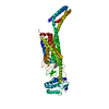
| ||||||||
|---|---|---|---|---|---|---|---|---|---|
| 1 |
| ||||||||
| Unit cell |
|
-Components
| #1: Protein | Mass: 68984.305 Da / Num. of mol.: 1 Source method: isolated from a genetically manipulated source Source: (gene. exp.) Gene: smo, Smo, cybC / Production host:   Spodoptera frugiperda (fall armyworm) Spodoptera frugiperda (fall armyworm)References: UniProt: Q98SW5, UniProt: P0ABE7, UniProt: A0A1L8GTP2*PLUS | ||
|---|---|---|---|
| #2: Chemical |   Mass: 411.620 Da / Num. of mol.: 2 / Source method: obtained synthetically / Formula: C27H41NO2 Mass: 411.620 Da / Num. of mol.: 2 / Source method: obtained synthetically / Formula: C27H41NO2Has protein modification | Y | |
-Experimental details
-Experiment
| Experiment | Method: X-RAY DIFFRACTION / Number of used crystals: 1 |
|---|
- Sample preparation
| Crystal | Density Matthews: 4.13 Å3/Da / Density % sol: 70.23 % |
|---|---|
| Crystal grow | Temperature: 293 K / Method: lipidic cubic phase Details: Reconstituted in 10:1 monoolein:cholesterol mix. Precipitant solution: 35-45% PEG 300, 300-500 mM LiSO4, 0.1 M MES pH 6-6.5 |
-Data collection
| Diffraction | Mean temperature: 80 K |
|---|---|
| Diffraction source | Source: SYNCHROTRON / Site: APS / Beamline: 23-ID-B / Wavelength: 1.0333 Å |
| Detector | Type: DECTRIS EIGER X 16M / Detector: PIXEL / Date: Oct 5, 2017 |
| Radiation | Protocol: SINGLE WAVELENGTH / Monochromatic (M) / Laue (L): M / Scattering type: x-ray |
| Radiation wavelength | Wavelength: 1.0333 Å / Relative weight: 1 |
| Reflection | Resolution: 3.74→49.63 Å / Num. obs: 11676 / % possible obs: 99.3 % / Redundancy: 6.5 % / CC1/2: 0.992 / Net I/σ(I): 6.1 |
| Reflection shell | Resolution: 3.74→3.81 Å / Redundancy: 5.6 % / Mean I/σ(I) obs: 1.9 / Num. unique obs: 504 / CC1/2: 0.617 / % possible all: 88.6 |
- Processing
| Software |
| |||||||||||||||||||||||||||||||||||
|---|---|---|---|---|---|---|---|---|---|---|---|---|---|---|---|---|---|---|---|---|---|---|---|---|---|---|---|---|---|---|---|---|---|---|---|---|
| Refinement | Method to determine structure: MOLECULAR REPLACEMENT Starting model: 5L7D, 5KZY and 4O9R Resolution: 3.751→41.054 Å / SU ML: 0.58 / Cross valid method: THROUGHOUT / σ(F): 0.31 / Phase error: 31.11
| |||||||||||||||||||||||||||||||||||
| Solvent computation | Shrinkage radii: 0.9 Å / VDW probe radii: 1.11 Å | |||||||||||||||||||||||||||||||||||
| Displacement parameters | Biso max: 195.84 Å2 / Biso mean: 90.0449 Å2 / Biso min: 40.22 Å2 | |||||||||||||||||||||||||||||||||||
| Refinement step | Cycle: final / Resolution: 3.751→41.054 Å
| |||||||||||||||||||||||||||||||||||
| Refine LS restraints |
| |||||||||||||||||||||||||||||||||||
| LS refinement shell | Refine-ID: X-RAY DIFFRACTION / Rfactor Rfree error: 0 / Total num. of bins used: 4
|
