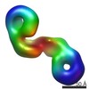
 Movie
Movie Controller

[English] 日本語
 Yorodumi
- PDB-4l8j: Crystal structure of a Putative efflux transporter (BACEGG_01895)... -
+ Open data

- Basic information
| Entry | Database: PDB / ID: 4l8j | ||||||
|---|---|---|---|---|---|---|---|
| Title | Crystal structure of a Putative efflux transporter (BACEGG_01895) from Bacteroides eggerthii DSM 20697 at 2.06 A resolution | ||||||
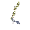
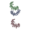
 Components Components | Putative efflux transporter | ||||||
 Keywords Keywords | TRANSPORT PROTEIN / HlyD family secretion protein / PF00529 family / Structural Genomics / Joint Center for Structural Genomics / JCSG / Protein Structure Initiative / PSI-BIOLOGY | ||||||
| Function / homology |  Function and homology information Function and homology informationconserved putative lor/sdh protein from methanococcus maripaludis s2 fold - #20 / conserved putative lor/sdh protein from methanococcus maripaludis s2 fold / Efflux pump adaptor protein, beta barrel domain / Helix hairpin bin / RNA polymerase II/Efflux pump adaptor protein, barrel-sandwich hybrid domain / Elongation Factor Tu (Ef-tu); domain 3 / OB fold (Dihydrolipoamide Acetyltransferase, E2P) / Helix Hairpins / Beta Barrel / Orthogonal Bundle ...conserved putative lor/sdh protein from methanococcus maripaludis s2 fold - #20 / conserved putative lor/sdh protein from methanococcus maripaludis s2 fold / Efflux pump adaptor protein, beta barrel domain / Helix hairpin bin / RNA polymerase II/Efflux pump adaptor protein, barrel-sandwich hybrid domain / Elongation Factor Tu (Ef-tu); domain 3 / OB fold (Dihydrolipoamide Acetyltransferase, E2P) / Helix Hairpins / Beta Barrel / Orthogonal Bundle / Mainly Beta / Mainly Alpha Similarity search - Domain/homology | ||||||
| Biological species |  Bacteroides eggerthii (bacteria) Bacteroides eggerthii (bacteria) | ||||||
| Method |  X-RAY DIFFRACTION / SYNCHROTRON / MAD / Resolution: 2.06 Å X-RAY DIFFRACTION / SYNCHROTRON / MAD / Resolution: 2.06 Å | ||||||
 Authors Authors | Joint Center for Structural Genomics (JCSG) | ||||||
 Citation Citation | Journal: To be published Title: Crystal structure of a Putative efflux transporter (BACEGG_01895) from Bacteroides eggerthii DSM 20697 at 2.06 A resolution Authors: Joint Center for Structural Genomics (JCSG) | ||||||
| History |
|
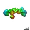
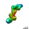
- Structure visualization
| Structure viewer | Molecule: MolmilJmol/JSmol |
|---|
- Downloads & links
-Download
| PDBx/mmCIF format | 4l8j.cif.gz | 148.5 KB | Display | PDBx/mmCIF format |
|---|---|---|---|---|
| PDB format | pdb4l8j.ent.gz | 115.4 KB | Display | PDB format |
| PDBx/mmJSON format | 4l8j.json.gz | Tree view | PDBx/mmJSON format | |
| Others |  Other downloads Other downloads |
-Validation report
| Arichive directory | https://data.pdbj.org/pub/pdb/validation_reports/l8/4l8jftp://data.pdbj.org/pub/pdb/validation_reports/l8/4l8j | HTTPS FTP |
|---|
-Related structure data
| Similar structure data | |
|---|---|
| Other databases |

-Links
 PDBj
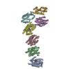
PDBj- Assembly
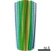
| Deposited unit | 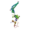
| ||||||||
|---|---|---|---|---|---|---|---|---|---|
| 1 | 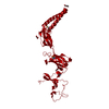
| ||||||||
| Unit cell |
| ||||||||
| Components on special symmetry positions |
|
-Components
| #1: Protein | Mass: 36749.359 Da / Num. of mol.: 1 Source method: isolated from a genetically manipulated source Source: (gene. exp.) Bacteroides eggerthii (bacteria) / Strain: DSM 20697 / Gene: BACEGG_01895, ZP_03459111.1 / Plasmid: SpeedET / Production host: | ||||||||
|---|---|---|---|---|---|---|---|---|---|
| #2: Chemical |   Mass: 106.120 Da / Num. of mol.: 3 / Source method: obtained synthetically / Formula: C4H10O3 Mass: 106.120 Da / Num. of mol.: 3 / Source method: obtained synthetically / Formula: C4H10O3#3: Chemical |   Mass: 92.094 Da / Num. of mol.: 2 / Source method: obtained synthetically / Formula: C3H8O3 Mass: 92.094 Da / Num. of mol.: 2 / Source method: obtained synthetically / Formula: C3H8O3#4: Water | ChemComp-HOH / |  Mass: 18.015 Da / Num. of mol.: 174 / Source method: isolated from a natural source / Formula: H2O Mass: 18.015 Da / Num. of mol.: 174 / Source method: isolated from a natural source / Formula: H2OHas protein modification | Y | Sequence details | THE CONSTRUCT (RESIDUES 24-350) WAS EXPRESSED WITH A PURIFICATION TAG MGSDKIHHHHHHENLYFQG. THE TAG ...THE CONSTRUCT (RESIDUES 24-350) WAS EXPRESSED WITH A PURIFICATI | |
-Experimental details
-Experiment
| Experiment | Method: X-RAY DIFFRACTION / Number of used crystals: 1 |
|---|
- Sample preparation
| Crystal | Density Matthews: 3.09 Å3/Da / Density % sol: 60.15 % |
|---|---|
| Crystal grow | Temperature: 277 K / Method: vapor diffusion, sitting drop / pH: 9.83 Details: 12.0% polyethylene glycol 8000, 0.1M sodium chloride, 0.1M CAPS pH 9.83, NANODROP, VAPOR DIFFUSION, SITTING DROP, temperature 277K |
-Data collection
| Diffraction | Mean temperature: 100 K | |||||||||||||||||||||||||||||||||||||||||||||||||||||||||||||||||||||||||||||
|---|---|---|---|---|---|---|---|---|---|---|---|---|---|---|---|---|---|---|---|---|---|---|---|---|---|---|---|---|---|---|---|---|---|---|---|---|---|---|---|---|---|---|---|---|---|---|---|---|---|---|---|---|---|---|---|---|---|---|---|---|---|---|---|---|---|---|---|---|---|---|---|---|---|---|---|---|---|---|
| Diffraction source | Source: SYNCHROTRON / Site: SSRL  / Beamline: BL11-1 / Wavelength: 0.97871, 0.91837, 0.97817 / Beamline: BL11-1 / Wavelength: 0.97871, 0.91837, 0.97817 | |||||||||||||||||||||||||||||||||||||||||||||||||||||||||||||||||||||||||||||
| Detector | Type: DECTRIS PILATUS 6M / Detector: PIXEL / Date: May 2, 2013 Details: Flat mirror (vertical focusing); single crystal Si(111) bent monochromator (horizontal focusing) | |||||||||||||||||||||||||||||||||||||||||||||||||||||||||||||||||||||||||||||
| Radiation | Monochromator: single crystal Si(111) bent / Protocol: MAD / Monochromatic (M) / Laue (L): M / Scattering type: x-ray | |||||||||||||||||||||||||||||||||||||||||||||||||||||||||||||||||||||||||||||
| Radiation wavelength |
| |||||||||||||||||||||||||||||||||||||||||||||||||||||||||||||||||||||||||||||
| Reflection | Resolution: 2.06→46.486 Å / Num. obs: 29410 / % possible obs: 99.8 % / Observed criterion σ(I): -3 / Biso Wilson estimate: 37.746 Å2 / Rmerge(I) obs: 0.076 / Net I/σ(I): 14.77 | |||||||||||||||||||||||||||||||||||||||||||||||||||||||||||||||||||||||||||||
| Reflection shell | Diffraction-ID: 1
|
-Phasing
| Phasing | Method: MAD |
|---|
- Processing
| Software |
| ||||||||||||||||||||||||||||||||||||||||||||||||||||||||||||||||||||||||||||||||||||||||||||||||||||||||||||
|---|---|---|---|---|---|---|---|---|---|---|---|---|---|---|---|---|---|---|---|---|---|---|---|---|---|---|---|---|---|---|---|---|---|---|---|---|---|---|---|---|---|---|---|---|---|---|---|---|---|---|---|---|---|---|---|---|---|---|---|---|---|---|---|---|---|---|---|---|---|---|---|---|---|---|---|---|---|---|---|---|---|---|---|---|---|---|---|---|---|---|---|---|---|---|---|---|---|---|---|---|---|---|---|---|---|---|---|---|---|
| Refinement | Method to determine structure: MAD / Resolution: 2.06→46.486 Å / Cor.coef. Fo:Fc: 0.9367 / Cor.coef. Fo:Fc free: 0.9229 / Occupancy max: 1 / Occupancy min: 0.5 / Cross valid method: THROUGHOUT / σ(F): 0 Details: 1. ATOM RECORD CONTAINS SUM OF TLS AND RESIDUAL B FACTORS. ANISOU RECORD CONTAINS SUM OF TLS AND RESIDUAL U FACTORS. 2. PEG FRAGMENTS (PEG) FROM THE CRYSTALLIZATION AND GLYCEROL (GOL) USED ...Details: 1. ATOM RECORD CONTAINS SUM OF TLS AND RESIDUAL B FACTORS. ANISOU RECORD CONTAINS SUM OF TLS AND RESIDUAL U FACTORS. 2. PEG FRAGMENTS (PEG) FROM THE CRYSTALLIZATION AND GLYCEROL (GOL) USED AS A CRYOPROTECTANT HAVE BEEN MODELED INTO THE STRUCTURE.3. THE REFINEMEMT WAS RESTRAINED AGAINST THE MAD PHASES.
| ||||||||||||||||||||||||||||||||||||||||||||||||||||||||||||||||||||||||||||||||||||||||||||||||||||||||||||
| Displacement parameters | Biso max: 116.81 Å2 / Biso mean: 46.9515 Å2 / Biso min: 24.58 Å2
| ||||||||||||||||||||||||||||||||||||||||||||||||||||||||||||||||||||||||||||||||||||||||||||||||||||||||||||
| Refine analyze | Luzzati coordinate error obs: 0.339 Å | ||||||||||||||||||||||||||||||||||||||||||||||||||||||||||||||||||||||||||||||||||||||||||||||||||||||||||||
| Refinement step | Cycle: LAST / Resolution: 2.06→46.486 Å
| ||||||||||||||||||||||||||||||||||||||||||||||||||||||||||||||||||||||||||||||||||||||||||||||||||||||||||||
| Refine LS restraints |
| ||||||||||||||||||||||||||||||||||||||||||||||||||||||||||||||||||||||||||||||||||||||||||||||||||||||||||||
| LS refinement shell | Resolution: 2.06→2.13 Å / Total num. of bins used: 15
| ||||||||||||||||||||||||||||||||||||||||||||||||||||||||||||||||||||||||||||||||||||||||||||||||||||||||||||
| Refinement TLS params. | Method: refined / Origin x: 21.2691 Å / Origin y: 10.4219 Å / Origin z: 54.4325 Å
| ||||||||||||||||||||||||||||||||||||||||||||||||||||||||||||||||||||||||||||||||||||||||||||||||||||||||||||
| Refinement TLS group | Selection details: { A|25 - 350 } |
