 Movie
Movie Controller


+ Open data

- Basic information
| Entry | Database: PDB / ID: 6avl | |||||||||
|---|---|---|---|---|---|---|---|---|---|---|
| Title | Orthorhombic Trypsin (295 K) in the presence of 50% xylose | |||||||||
 Components Components | Cationic trypsin | |||||||||
 Keywords Keywords | HYDROLASE / serine protease | |||||||||
| Function / homology |  Function and homology information Function and homology informationtrypsin / serpin family protein binding / serine protease inhibitor complex / digestion / endopeptidase activity / serine-type endopeptidase activity / proteolysis / extracellular space / metal ion binding Similarity search - Function | |||||||||
| Biological species |  | |||||||||
| Method |  X-RAY DIFFRACTION / MOLECULAR REPLACEMENT / Resolution: 2 Å X-RAY DIFFRACTION / MOLECULAR REPLACEMENT / Resolution: 2 Å | |||||||||
 Authors Authors | Juers, D.H. | |||||||||
| Funding support |  United States, 1items United States, 1items
| |||||||||
 Citation Citation | Journal: Acta Crystallogr D Struct Biol / Year: 2018 Title: The impact of cryosolution thermal contraction on proteins and protein crystals: volumes, conformation and order. Authors: Juers, D.H. / Farley, C.A. / Saxby, C.P. / Cotter, R.A. / Cahn, J.K.B. / Holton-Burke, R.C. / Harrison, K. / Wu, Z. | |||||||||
| History |
|
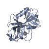
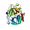
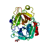
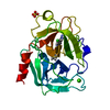
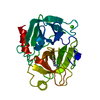
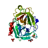
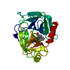
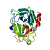
- Structure visualization
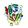
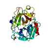
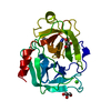
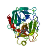
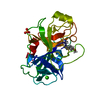
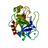
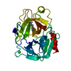
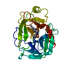
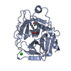
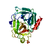
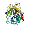
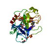
| Structure viewer | Molecule: MolmilJmol/JSmol |
|---|
- Downloads & links
-Download
| PDBx/mmCIF format | 6avl.cif.gz | 98.3 KB | Display | PDBx/mmCIF format |
|---|---|---|---|---|
| PDB format | pdb6avl.ent.gz | 73.4 KB | Display | PDB format |
| PDBx/mmJSON format | 6avl.json.gz | Tree view | PDBx/mmJSON format | |
| Others |  Other downloads Other downloads |
-Validation report
| Summary document | 6avl_validation.pdf.gz | 458.3 KB | Display | wwPDB validaton report |
|---|---|---|---|---|
| Full document | 6avl_full_validation.pdf.gz | 458.2 KB | Display | |
| Data in XML | 6avl_validation.xml.gz | 13.7 KB | Display | |
| Data in CIF | 6avl_validation.cif.gz | 18.6 KB | Display | |
| Arichive directory | https://data.pdbj.org/pub/pdb/validation_reports/av/6avlftp://data.pdbj.org/pub/pdb/validation_reports/av/6avl | HTTPS FTP |
-Related structure data
| Related structure data | 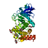 5un3C 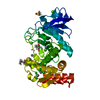 5uu7C 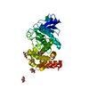 5uu8C  5uu9C 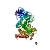 5uuaC 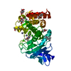 5uubC 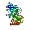 5uucC 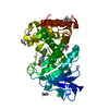 5uudC 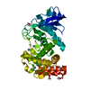 5uueC 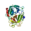 6b6nC 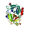 6b6oC 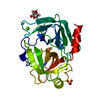 6b6pC 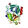 6b6qC 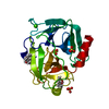 6b6rC  6b6sC 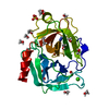 6b6tC  6d5nC 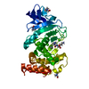 6d5oC 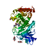 6d5pC 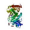 6d5qC 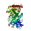 6d5rC 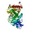 6d5sC 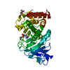 6d5tC 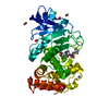 6d5uC 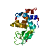 6d6eC 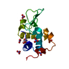 6d6fC 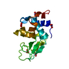 6d6gC 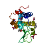 6d6hC 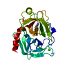 6dzfC C: citing same article ( |
|---|---|
| Similar structure data |
-Links
 PDBj
PDBj


- Assembly
| Deposited unit | 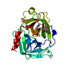
| ||||||||
|---|---|---|---|---|---|---|---|---|---|
| 1 |
| ||||||||
| Unit cell |
|
-Components
-Protein , 1 types, 1 molecules A
| #1: Protein | Mass: 23324.287 Da / Num. of mol.: 1 Source method: isolated from a genetically manipulated source Source: (gene. exp.) |
|---|
-Sugars , 2 types, 2 molecules 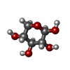
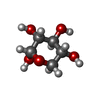
| #5: Sugar | ChemComp-XYP /  Type: D-saccharide, beta linking / Mass: 150.130 Da / Num. of mol.: 1 Type: D-saccharide, beta linking / Mass: 150.130 Da / Num. of mol.: 1Source method: isolated from a genetically manipulated source Formula: C5H10O5 |
|---|---|
| #6: Sugar | ChemComp-XYS /  Type: D-saccharide, alpha linking / Mass: 150.130 Da / Num. of mol.: 1 Type: D-saccharide, alpha linking / Mass: 150.130 Da / Num. of mol.: 1Source method: isolated from a genetically manipulated source Formula: C5H10O5 |
-Non-polymers , 4 types, 170 molecules 


| #2: Chemical | ChemComp-CA /  Mass: 40.078 Da / Num. of mol.: 1 / Source method: obtained synthetically / Formula: Ca Mass: 40.078 Da / Num. of mol.: 1 / Source method: obtained synthetically / Formula: Ca |
|---|---|
| #3: Chemical | ChemComp-BEN /  Mass: 120.152 Da / Num. of mol.: 1 / Source method: obtained synthetically / Formula: C7H8N2 Mass: 120.152 Da / Num. of mol.: 1 / Source method: obtained synthetically / Formula: C7H8N2 |
| #4: Chemical | ChemComp-SO4 /  Mass: 96.063 Da / Num. of mol.: 1 / Source method: obtained synthetically / Formula: SO4 Mass: 96.063 Da / Num. of mol.: 1 / Source method: obtained synthetically / Formula: SO4 |
| #7: Water | ChemComp-HOH / Mass: 18.015 Da / Num. of mol.: 167 / Source method: isolated from a natural source / Formula: H2O |
-Details
| Has protein modification | Y |
|---|
-Experimental details
-Experiment
| Experiment | Method: X-RAY DIFFRACTION / Number of used crystals: 1 |
|---|
- Sample preparation
| Crystal | Density Matthews: 2.33 Å3/Da / Density % sol: 47.25 % |
|---|---|
| Crystal grow | Temperature: 295 K / Method: vapor diffusion, hanging drop / pH: 8 Details: 0.1 M Tris buffer 0.2 M AmSO4 25% (w/v) PEG 8000 0.1 M benzamidine-HCl Protein 40 mg/mL in water |
-Data collection
| Diffraction | Mean temperature: 295 K |
|---|---|
| Diffraction source | Source: SEALED TUBE / Type: OXFORD DIFFRACTION NOVA / Wavelength: 1.54 Å |
| Detector | Type: OXFORD ONYX CCD / Detector: CCD / Date: Jun 1, 2017 |
| Radiation | Protocol: SINGLE WAVELENGTH / Monochromatic (M) / Laue (L): M / Scattering type: x-ray |
| Radiation wavelength | Wavelength: 1.54 Å / Relative weight: 1 |
| Reflection | Resolution: 2→13.79 Å / Num. obs: 15148 / % possible obs: 99.2 % / Redundancy: 3.7 % / CC1/2: 0.98 / Net I/σ(I): 5.4 |
| Reflection shell | Resolution: 2→2.11 Å / CC1/2: 0.87 |
- Processing
| Software |
| |||||||||||||||||||||||||||||||||||||||||||||||||
|---|---|---|---|---|---|---|---|---|---|---|---|---|---|---|---|---|---|---|---|---|---|---|---|---|---|---|---|---|---|---|---|---|---|---|---|---|---|---|---|---|---|---|---|---|---|---|---|---|---|---|
| Refinement | Method to determine structure: MOLECULAR REPLACEMENT / Resolution: 2→13.468 Å / SU ML: 0.2 / Cross valid method: FREE R-VALUE / σ(F): 1.35 / Phase error: 19.96
| |||||||||||||||||||||||||||||||||||||||||||||||||
| Solvent computation | Shrinkage radii: 0.9 Å / VDW probe radii: 1.11 Å | |||||||||||||||||||||||||||||||||||||||||||||||||
| Refinement step | Cycle: LAST / Resolution: 2→13.468 Å
| |||||||||||||||||||||||||||||||||||||||||||||||||
| Refine LS restraints |
| |||||||||||||||||||||||||||||||||||||||||||||||||
| LS refinement shell |
|
