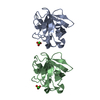
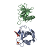
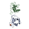
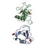
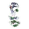
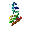
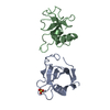
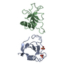
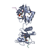
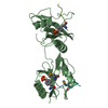
Entry Database : PDB / ID : 5x94Title Crystal structure of SHP2_SH2-CagA EPIYA_D peptide complex Cag pathogenicity island protein Tyrosine-protein phosphatase non-receptor type 11 Keywords / / / Function / homology Function Domain/homology Component
/ / / / / / / / / / / / / / / / / / / / / / / / / / / / / / / / / / / / / / / / / / / / / / / / / / / / / / / / / / / / / / / / / / / / / / / / / / / / / / / / / / / / / / / / / / / / / / / / / / / / / / / / / / / / / / / / / / / / / / / / / / / / / / / / / / / / / / / / / / / / / / / / Biological species Homo sapiens (human)Helicobacter pylori (bacteria)Method / / / Resolution : 2.6 Å Authors Senda, M. / Senda, T. Journal : Cell Rep / Year : 2017Title : Differential Mechanisms for SHP2 Binding and Activation Are Exploited by Geographically Distinct Helicobacter pylori CagA Oncoproteins.Authors : Hayashi, T. / Senda, M. / Suzuki, N. / Nishikawa, H. / Ben, C. / Tang, C. / Nagase, L. / Inoue, K. / Senda, T. / Hatakeyama, M. History Deposition Mar 5, 2017 Deposition site / Processing site Revision 1.0 Sep 13, 2017 Provider / Type Revision 1.1 Oct 18, 2017 Group / Category / citation_authorItem _citation.country / _citation.journal_abbrev ... _citation.country / _citation.journal_abbrev / _citation.journal_id_CSD / _citation.journal_id_ISSN / _citation.journal_volume / _citation.page_first / _citation.page_last / _citation.pdbx_database_id_DOI / _citation.pdbx_database_id_PubMed / _citation.title / _citation.year Revision 1.2 Nov 22, 2023 Group / Database references / Refinement descriptionCategory chem_comp_atom / chem_comp_bond ... chem_comp_atom / chem_comp_bond / database_2 / pdbx_initial_refinement_model Item / _database_2.pdbx_database_accessionRevision 1.3 Oct 23, 2024 Group / Category / pdbx_modification_feature
Show all Show less
 Movie
Movie Controller


 Open data
 Basic information
 Components
 Keywords
 Function and homology information
 Homo sapiens (human)
 Helicobacter pylori (bacteria)
 X-RAY DIFFRACTION /
  Authors
 Citation
 Structure visualization
 Downloads & links
 Other downloads
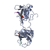


 PDBj
PDBj


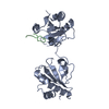
 Assembly
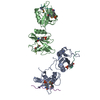
 Mass: 18.015 Da / Num. of mol.: 22 / Source method: isolated from a natural source / Formula: H2O
 Sample preparation
 / Beamline: BL-5A / Wavelength: 0.979 Å
 Processing