 Movie
Movie Controller

[English] 日本語
 Yorodumi
- PDB-4j5k: Crystal structure analysis of Streptomyces aureofaciens ribonucle... -
+ Open data

- Basic information
| Entry | Database: PDB / ID: 4j5k | ||||||
|---|---|---|---|---|---|---|---|
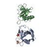
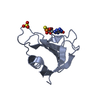
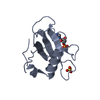
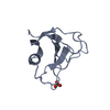
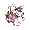
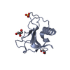
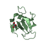
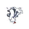
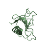
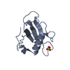
| Title | Crystal structure analysis of Streptomyces aureofaciens ribonuclease Sa Y51F mutant | ||||||
 Components Components | Guanyl-specific ribonuclease Sa | ||||||
 Keywords Keywords | HYDROLASE / endoribonuclease / mutant | ||||||
| Function / homology |  Function and homology information Function and homology informationribonuclease T1 / ribonuclease T1 activity / hydrolase activity / RNA binding / extracellular region Similarity search - Function | ||||||
| Biological species |  Streptomyces aureofaciens (bacteria) Streptomyces aureofaciens (bacteria) | ||||||
| Method |  X-RAY DIFFRACTION / SYNCHROTRON / Resolution: 1.23 Å X-RAY DIFFRACTION / SYNCHROTRON / Resolution: 1.23 Å | ||||||
 Authors Authors | Urbanikova, L. / Sevcik, J. | ||||||
 Citation Citation | Journal: Protein Sci. / Year: 2014 Title: Contribution of hydrogen bonds to protein stability. Authors: Pace, C.N. / Fu, H. / Lee Fryar, K. / Landua, J. / Trevino, S.R. / Schell, D. / Thurlkill, R.L. / Imura, S. / Scholtz, J.M. / Gajiwala, K. / Sevcik, J. / Urbanikova, L. / Myers, J.K. / ...Authors: Pace, C.N. / Fu, H. / Lee Fryar, K. / Landua, J. / Trevino, S.R. / Schell, D. / Thurlkill, R.L. / Imura, S. / Scholtz, J.M. / Gajiwala, K. / Sevcik, J. / Urbanikova, L. / Myers, J.K. / Takano, K. / Hebert, E.J. / Shirley, B.A. / Grimsley, G.R. | ||||||
| History |
|
- Structure visualization
| Structure viewer | Molecule: MolmilJmol/JSmol |
|---|
- Downloads & links
-Download
| PDBx/mmCIF format | 4j5k.cif.gz | 111.9 KB | Display | PDBx/mmCIF format |
|---|---|---|---|---|
| PDB format | pdb4j5k.ent.gz | 88.6 KB | Display | PDB format |
| PDBx/mmJSON format | 4j5k.json.gz | Tree view | PDBx/mmJSON format | |
| Others |  Other downloads Other downloads |
-Validation report
| Arichive directory | https://data.pdbj.org/pub/pdb/validation_reports/j5/4j5kftp://data.pdbj.org/pub/pdb/validation_reports/j5/4j5k | HTTPS FTP |
|---|
-Related structure data
| Related structure data |  4ghoC  4j5gC  1t2hS C: citing same article ( S: Starting model for refinement |
|---|---|
| Similar structure data |


-Links
 PDBj
PDBj- Assembly
| Deposited unit | 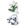
| ||||||||
|---|---|---|---|---|---|---|---|---|---|
| 1 | 
| ||||||||
| 2 | 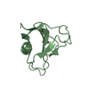
| ||||||||
| 3 | 
| ||||||||
| Unit cell |
|
-Components
| #1: Protein | Mass: 10566.492 Da / Num. of mol.: 2 / Fragment: Ribonuclease Sa / Mutation: Y51F Source method: isolated from a genetically manipulated source Source: (gene. exp.) Streptomyces aureofaciens (bacteria) / Strain: BMK / Gene: rnaSA / Production host: #2: Chemical | ChemComp-SO4 / |   Mass: 96.063 Da / Num. of mol.: 1 / Source method: obtained synthetically / Formula: SO4 Mass: 96.063 Da / Num. of mol.: 1 / Source method: obtained synthetically / Formula: SO4#3: Chemical |   Mass: 92.094 Da / Num. of mol.: 2 / Source method: obtained synthetically / Formula: C3H8O3 Mass: 92.094 Da / Num. of mol.: 2 / Source method: obtained synthetically / Formula: C3H8O3#4: Water | ChemComp-HOH / |  Mass: 18.015 Da / Num. of mol.: 416 / Source method: isolated from a natural source / Formula: H2O Mass: 18.015 Da / Num. of mol.: 416 / Source method: isolated from a natural source / Formula: H2OHas protein modification | Y | |
|---|
-Experimental details
-Experiment
| Experiment | Method: X-RAY DIFFRACTION / Number of used crystals: 1 |
|---|
- Sample preparation
| Crystal | Density Matthews: 2.24 Å3/Da / Density % sol: 45.2 % |
|---|---|
| Crystal grow | Temperature: 293 K / Method: vapor diffusion, hanging drop / pH: 7.4 Details: Ammonium sulfate 0.9 M, phosphate buffer 0.1 M , pH 7.4, VAPOR DIFFUSION, HANGING DROP, temperature 293K |
-Data collection
| Diffraction | Mean temperature: 100 K |
|---|---|
| Diffraction source | Source: SYNCHROTRON / Site: EMBL/DESY, HAMBURG  / Beamline: X31 / Wavelength: 1.1 Å / Beamline: X31 / Wavelength: 1.1 Å |
| Detector | Type: MAR scanner 345 mm plate / Detector: IMAGE PLATE / Date: Jul 9, 2002 / Details: mirrors |
| Radiation | Monochromator: Si 111 CHANNEL / Protocol: SINGLE WAVELENGTH / Monochromatic (M) / Laue (L): M / Scattering type: x-ray |
| Radiation wavelength | Wavelength: 1.1 Å / Relative weight: 1 |
| Reflection | Resolution: 1.23→20 Å / Num. all: 56065 / Num. obs: 54845 / % possible obs: 97.8 % / Redundancy: 2.3 % / Rmerge(I) obs: 0.037 / Net I/σ(I): 29.7 |
| Reflection shell | Resolution: 1.23→1.24 Å / Redundancy: 1.8 % / Rmerge(I) obs: 0.139 / Mean I/σ(I) obs: 4.9 / Num. unique all: 1493 / % possible all: 82.6 |
- Processing
| Software |
| |||||||||||||||||||||||||||||||||||||||||||||||||||||||||||||||||||||||||||
|---|---|---|---|---|---|---|---|---|---|---|---|---|---|---|---|---|---|---|---|---|---|---|---|---|---|---|---|---|---|---|---|---|---|---|---|---|---|---|---|---|---|---|---|---|---|---|---|---|---|---|---|---|---|---|---|---|---|---|---|---|---|---|---|---|---|---|---|---|---|---|---|---|---|---|---|---|
| Refinement | Starting model: 1T2H Resolution: 1.23→18.59 Å / Cor.coef. Fo:Fc: 0.984 / Cor.coef. Fo:Fc free: 0.979 / SU B: 1.085 / SU ML: 0.022 / Cross valid method: THROUGHOUT / ESU R: 0.033 / ESU R Free: 0.034 / Stereochemistry target values: MAXIMUM LIKELIHOOD / Details: HYDROGENS HAVE BEEN ADDED IN THE RIDING POSITIONS
| |||||||||||||||||||||||||||||||||||||||||||||||||||||||||||||||||||||||||||
| Solvent computation | Ion probe radii: 0.8 Å / Shrinkage radii: 0.8 Å / VDW probe radii: 1.2 Å / Solvent model: MASK | |||||||||||||||||||||||||||||||||||||||||||||||||||||||||||||||||||||||||||
| Displacement parameters | Biso mean: 11.99 Å2
| |||||||||||||||||||||||||||||||||||||||||||||||||||||||||||||||||||||||||||
| Refinement step | Cycle: LAST / Resolution: 1.23→18.59 Å
| |||||||||||||||||||||||||||||||||||||||||||||||||||||||||||||||||||||||||||
| Refine LS restraints |
| |||||||||||||||||||||||||||||||||||||||||||||||||||||||||||||||||||||||||||
| LS refinement shell | Resolution: 1.23→1.262 Å / Total num. of bins used: 20
|
