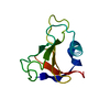
 Movie
Movie Controller


+ Open data

- Basic information
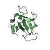
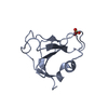
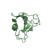
| Entry | Database: PDB / ID: 1i8v | ||||||
|---|---|---|---|---|---|---|---|
| Title | CRYSTAL STRUCTURE OF RNASE SA Y80F MUTANT | ||||||
 Components Components | GUANYL-SPECIFIC RIBONUCLEASE SA | ||||||
 Keywords Keywords | HYDROLASE / mutant | ||||||
| Function / homology |  Function and homology information Function and homology informationribonuclease T1 / ribonuclease T1 activity / hydrolase activity / RNA binding / extracellular region Similarity search - Function | ||||||
| Biological species |  Streptomyces aureofaciens (bacteria) Streptomyces aureofaciens (bacteria) | ||||||
| Method |  X-RAY DIFFRACTION / SYNCHROTRON / STARTING MODEL WAS USED WITHOUT MOLECULAR REPLACEMENT / Resolution: 1.25 Å X-RAY DIFFRACTION / SYNCHROTRON / STARTING MODEL WAS USED WITHOUT MOLECULAR REPLACEMENT / Resolution: 1.25 Å | ||||||
 Authors Authors | Sevcik, J. / Urbanikova, L. | ||||||
 Citation Citation | Journal: J.Mol.Biol. / Year: 2001 Title: Tyrosine hydrogen bonds make a large contribution to protein stability. Authors: Pace, C.N. / Horn, G. / Hebert, E.J. / Bechert, J. / Shaw, K. / Urbanikova, L. / Scholtz, J.M. / Sevcik, J. | ||||||
| History |
|
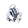
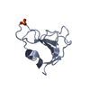
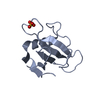
- Structure visualization
| Structure viewer | Molecule: MolmilJmol/JSmol |
|---|
- Downloads & links
-Download
| PDBx/mmCIF format | 1i8v.cif.gz | 56.2 KB | Display | PDBx/mmCIF format |
|---|---|---|---|---|
| PDB format | pdb1i8v.ent.gz | 40.6 KB | Display | PDB format |
| PDBx/mmJSON format | 1i8v.json.gz | Tree view | PDBx/mmJSON format | |
| Others |  Other downloads Other downloads |
-Validation report
| Arichive directory | https://data.pdbj.org/pub/pdb/validation_reports/i8/1i8vftp://data.pdbj.org/pub/pdb/validation_reports/i8/1i8v | HTTPS FTP |
|---|
-Related structure data
| Related structure data |  1i70C 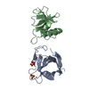 1rggS  1i8w S: Starting model for refinement C: citing same article ( |
|---|---|
| Similar structure data |


-Links
 PDBj
PDBj- Assembly
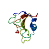
| Deposited unit | 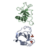
| ||||||||
|---|---|---|---|---|---|---|---|---|---|
| 1 | 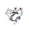
| ||||||||
| 2 | 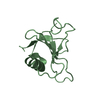
| ||||||||
| Unit cell |
|
-Components
| #1: Protein | Mass: 10566.492 Da / Num. of mol.: 2 / Mutation: Y80F Source method: isolated from a genetically manipulated source Source: (gene. exp.) Streptomyces aureofaciens (bacteria) / Production host: #2: Chemical |   Mass: 96.063 Da / Num. of mol.: 2 / Source method: obtained synthetically / Formula: SO4 Mass: 96.063 Da / Num. of mol.: 2 / Source method: obtained synthetically / Formula: SO4#3: Water | ChemComp-HOH / |  Mass: 18.015 Da / Num. of mol.: 287 / Source method: isolated from a natural source / Formula: H2O Mass: 18.015 Da / Num. of mol.: 287 / Source method: isolated from a natural source / Formula: H2OHas protein modification | Y | |
|---|
-Experimental details
-Experiment
| Experiment | Method: X-RAY DIFFRACTION / Number of used crystals: 1 |
|---|
- Sample preparation
| Crystal | Density Matthews: 2.35 Å3/Da / Density % sol: 47.7 % | ||||||||||||||||||||
|---|---|---|---|---|---|---|---|---|---|---|---|---|---|---|---|---|---|---|---|---|---|
| Crystal grow | Temperature: 293 K / Method: vapor diffusion, hanging drop / pH: 7 Details: AMMONIUM SULFATE, MONOSODIUM PHOSPHATE, DISODIUM PHOSPHATE, pH 7.0, VAPOR DIFFUSION, HANGING DROP, temperature 293.0K | ||||||||||||||||||||
| Crystal grow | *PLUS Details: used microseeding / PH range low: 7.2 / PH range high: 7 | ||||||||||||||||||||
| Components of the solutions | *PLUS
|
-Data collection
| Diffraction | Mean temperature: 293 K |
|---|---|
| Diffraction source | Source: SYNCHROTRON / Site: EMBL/DESY, HAMBURG  / Beamline: X31 / Wavelength: 1.044 Å / Beamline: X31 / Wavelength: 1.044 Å |
| Detector | Type: MARRESEARCH / Detector: IMAGE PLATE / Date: Nov 1, 1999 |
| Radiation | Monochromator: SI 111 CHANNEL-CUT XTAL / Protocol: SINGLE WAVELENGTH / Monochromatic (M) / Laue (L): M / Scattering type: x-ray |
| Radiation wavelength | Wavelength: 1.044 Å / Relative weight: 1 |
| Reflection | Resolution: 1.25→19.65 Å / Num. all: 52680 / Num. obs: 52680 / % possible obs: 94.3 % / Redundancy: 6.1 % / Biso Wilson estimate: 13.391 Å2 / Rmerge(I) obs: 0.057 / Net I/σ(I): 29.09 |
| Reflection shell | Resolution: 1.25→1.26 Å / Rmerge(I) obs: 0.426 / Mean I/σ(I) obs: 2.28 / % possible all: 83.5 |
| Reflection | *PLUS |
- Processing
| Software |
| ||||||||||||||||||||||||||||||||||||||||||||||||||||||||||||||||||||||||||||||||||||
|---|---|---|---|---|---|---|---|---|---|---|---|---|---|---|---|---|---|---|---|---|---|---|---|---|---|---|---|---|---|---|---|---|---|---|---|---|---|---|---|---|---|---|---|---|---|---|---|---|---|---|---|---|---|---|---|---|---|---|---|---|---|---|---|---|---|---|---|---|---|---|---|---|---|---|---|---|---|---|---|---|---|---|---|---|---|
| Refinement | Method to determine structure: STARTING MODEL WAS USED WITHOUT MOLECULAR REPLACEMENT Starting model: PDB ENTRY 1RGG Resolution: 1.25→19.6 Å / SU B: 0.717 / SU ML: 0.03 / ESU R: 0.037 / ESU R Free: 0.038 / Stereochemistry target values: ENGH & HUBER
| ||||||||||||||||||||||||||||||||||||||||||||||||||||||||||||||||||||||||||||||||||||
| Refinement step | Cycle: LAST / Resolution: 1.25→19.6 Å
| ||||||||||||||||||||||||||||||||||||||||||||||||||||||||||||||||||||||||||||||||||||
| Refine LS restraints |
| ||||||||||||||||||||||||||||||||||||||||||||||||||||||||||||||||||||||||||||||||||||
| LS refinement shell | Resolution: 1.25→1.26 Å | ||||||||||||||||||||||||||||||||||||||||||||||||||||||||||||||||||||||||||||||||||||
| Software | *PLUS Name: REFMAC / Classification: refinement | ||||||||||||||||||||||||||||||||||||||||||||||||||||||||||||||||||||||||||||||||||||
| Refinement | *PLUS % reflection Rfree: 5 % / Rfactor Rwork: 0.13 | ||||||||||||||||||||||||||||||||||||||||||||||||||||||||||||||||||||||||||||||||||||
| Solvent computation | *PLUS | ||||||||||||||||||||||||||||||||||||||||||||||||||||||||||||||||||||||||||||||||||||
| Displacement parameters | *PLUS |
