 Movie
Movie Controller


+ Open data

- Basic information
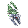
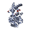
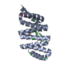
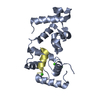
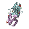
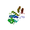
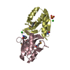
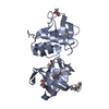
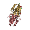
| Entry | Database: PDB / ID: 1ay7 | ||||||
|---|---|---|---|---|---|---|---|
| Title | RIBONUCLEASE SA COMPLEX WITH BARSTAR | ||||||
 Components Components |
| ||||||
 Keywords Keywords | COMPLEX (ENZYME/INHIBITOR) / RIBONUCLEASE / INHIBITOR / STREPTOMYCES AUREOFACIENS / COMPLEX (ENZYME-INHIBITOR) / COMPLEX (ENZYME-INHIBITOR) complex | ||||||
| Function / homology |  Function and homology information Function and homology informationribonuclease T1 / ribonuclease T1 activity / hydrolase activity / RNA binding / extracellular region / cytoplasm Similarity search - Function | ||||||
| Biological species |  Streptomyces aureofaciens (bacteria) Streptomyces aureofaciens (bacteria) | ||||||
| Method |  X-RAY DIFFRACTION / SYNCHROTRON / MOLECULAR REPLACEMENT / Resolution: 1.7 Å X-RAY DIFFRACTION / SYNCHROTRON / MOLECULAR REPLACEMENT / Resolution: 1.7 Å | ||||||
 Authors Authors | Sevcik, J. / Urbanikova, L. / Dauter, Z. / Wilson, K.S. | ||||||
 Citation Citation | Journal: Acta Crystallogr.,Sect.D / Year: 1998 Title: Recognition of RNase Sa by the inhibitor barstar: structure of the complex at 1.7 A resolution. Authors: Sevcik, J. / Urbanikova, L. / Dauter, Z. / Wilson, K.S. | ||||||
| History |
|
- Structure visualization
| Structure viewer | Molecule: MolmilJmol/JSmol |
|---|
- Downloads & links
-Download
| PDBx/mmCIF format | 1ay7.cif.gz | 54.1 KB | Display | PDBx/mmCIF format |
|---|---|---|---|---|
| PDB format | pdb1ay7.ent.gz | 38.9 KB | Display | PDB format |
| PDBx/mmJSON format | 1ay7.json.gz | Tree view | PDBx/mmJSON format | |
| Others |  Other downloads Other downloads |
-Validation report
| Arichive directory | https://data.pdbj.org/pub/pdb/validation_reports/ay/1ay7ftp://data.pdbj.org/pub/pdb/validation_reports/ay/1ay7 | HTTPS FTP |
|---|
-Related structure data
| Related structure data |  1brsS S: Starting model for refinement |
|---|---|
| Similar structure data |

-Links
 PDBj
PDBj- Assembly
| Deposited unit | 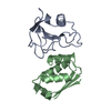
| ||||||||
|---|---|---|---|---|---|---|---|---|---|
| 1 |
| ||||||||
| Unit cell |
|
-Components
| #1: Protein | Mass: 10582.492 Da / Num. of mol.: 1 Source method: isolated from a genetically manipulated source Source: (gene. exp.) Streptomyces aureofaciens (bacteria) / Plasmid: PMT1126 / Production host: |
|---|---|
| #2: Protein | Mass: 10221.543 Da / Num. of mol.: 1 Source method: isolated from a genetically manipulated source Details: BARNASE INHIBITOR / Source: (gene. exp.) |
| #3: Water | ChemComp-HOH /  Mass: 18.015 Da / Num. of mol.: 190 / Source method: isolated from a natural source / Formula: H2O Mass: 18.015 Da / Num. of mol.: 190 / Source method: isolated from a natural source / Formula: H2O |
| Has protein modification | Y |
-Experimental details
-Experiment
| Experiment | Method: X-RAY DIFFRACTION / Number of used crystals: 1 |
|---|
- Sample preparation
| Crystal | Density Matthews: 3.35 Å3/Da / Density % sol: 69 % | |||||||||||||||||||||||||
|---|---|---|---|---|---|---|---|---|---|---|---|---|---|---|---|---|---|---|---|---|---|---|---|---|---|---|
| Crystal grow | pH: 7 / Details: pH 7.0 | |||||||||||||||||||||||||
| Crystal | *PLUS | |||||||||||||||||||||||||
| Crystal grow | *PLUS pH: 7.2 / Method: vapor diffusion | |||||||||||||||||||||||||
| Components of the solutions | *PLUS
|
-Data collection
| Diffraction | Mean temperature: 293 K |
|---|---|
| Diffraction source | Source: SYNCHROTRON / Site: EMBL/DESY, HAMBURG  / Beamline: X31 / Wavelength: 0.95 / Beamline: X31 / Wavelength: 0.95 |
| Detector | Type: MARRESEARCH / Detector: IMAGE PLATE / Date: Nov 1, 1996 / Details: MIRRORS |
| Radiation | Monochromator: SI(111) / Monochromatic (M) / Laue (L): M / Scattering type: x-ray |
| Radiation wavelength | Wavelength: 0.95 Å / Relative weight: 1 |
| Reflection | Resolution: 1.7→25 Å / Num. obs: 27413 / % possible obs: 100 % / Observed criterion σ(I): -3 / Redundancy: 4.7 % / Biso Wilson estimate: 20 Å2 / Rmerge(I) obs: 0.046 / Rsym value: 0.046 / Net I/σ(I): 27.3 |
| Reflection shell | Resolution: 1.7→1.72 Å / Redundancy: 4.2 % / Rmerge(I) obs: 0.308 / Mean I/σ(I) obs: 3.7 / Rsym value: 0.308 / % possible all: 99.8 |
| Reflection | *PLUS Num. all: 27413 / Num. obs: 22844 / Num. measured all: 118438 |
- Processing
| Software |
| ||||||||||||||||||||||||||||||||||||||||||||||||||||||||||||||||||||||||||||||||||||
|---|---|---|---|---|---|---|---|---|---|---|---|---|---|---|---|---|---|---|---|---|---|---|---|---|---|---|---|---|---|---|---|---|---|---|---|---|---|---|---|---|---|---|---|---|---|---|---|---|---|---|---|---|---|---|---|---|---|---|---|---|---|---|---|---|---|---|---|---|---|---|---|---|---|---|---|---|---|---|---|---|---|---|---|---|---|
| Refinement | Method to determine structure: MOLECULAR REPLACEMENT Starting model: PDB ENTRY 1BRS Resolution: 1.7→25 Å / Cross valid method: FREE R / σ(F): 0 Details: ESTIMATED COORDINATE ERROR. ESD FROM CRUICKSHANK (A) : 0.078 ESD FROM SIGMAA (A) : 0.061
| ||||||||||||||||||||||||||||||||||||||||||||||||||||||||||||||||||||||||||||||||||||
| Displacement parameters | Biso mean: 24.8 Å2
| ||||||||||||||||||||||||||||||||||||||||||||||||||||||||||||||||||||||||||||||||||||
| Refine analyze | Luzzati sigma a obs: 0.06 Å | ||||||||||||||||||||||||||||||||||||||||||||||||||||||||||||||||||||||||||||||||||||
| Refinement step | Cycle: LAST / Resolution: 1.7→25 Å
| ||||||||||||||||||||||||||||||||||||||||||||||||||||||||||||||||||||||||||||||||||||
| Refine LS restraints |
| ||||||||||||||||||||||||||||||||||||||||||||||||||||||||||||||||||||||||||||||||||||
| Software | *PLUS Name: REFMAC / Classification: refinement | ||||||||||||||||||||||||||||||||||||||||||||||||||||||||||||||||||||||||||||||||||||
| Refinement | *PLUS Rfactor Rfree: 0.19 / Rfactor Rwork: 0.16 | ||||||||||||||||||||||||||||||||||||||||||||||||||||||||||||||||||||||||||||||||||||
| Solvent computation | *PLUS | ||||||||||||||||||||||||||||||||||||||||||||||||||||||||||||||||||||||||||||||||||||
| Displacement parameters | *PLUS |
