 Movie
Movie Controller


+ Open data

- Basic information
| Entry | Database: PDB / ID: 1box | ||||||
|---|---|---|---|---|---|---|---|
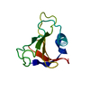
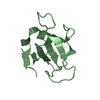
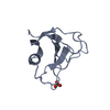
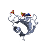
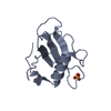
| Title | N39S MUTANT OF RNASE SA FROM STREPTOMYCES AUREOFACIENS | ||||||
 Components Components | GUANYL-SPECIFIC RIBONUCLEASE SA | ||||||
 Keywords Keywords | HYDROLASE / RIBONUCLEASE / MUTANT | ||||||
| Function / homology |  Function and homology information Function and homology informationribonuclease T1 / ribonuclease T1 activity / hydrolase activity / RNA binding / extracellular region Similarity search - Function | ||||||
| Biological species |  Streptomyces aureofaciens (bacteria) Streptomyces aureofaciens (bacteria) | ||||||
| Method |  X-RAY DIFFRACTION / MOLECULAR REPLACEMENT / Resolution: 1.6 Å X-RAY DIFFRACTION / MOLECULAR REPLACEMENT / Resolution: 1.6 Å | ||||||
 Authors Authors | Hebert, E.J. / Giletto, A. / Sevcik, J. / Urbanikova, L. / Wilson, K.S. / Dauter, Z. / Pace, C.N. | ||||||
 Citation Citation | Journal: Biochemistry / Year: 1998 Title: Contribution of a conserved asparagine to the conformational stability of ribonucleases Sa, Ba, and T1. Authors: Hebert, E.J. / Giletto, A. / Sevcik, J. / Urbanikova, L. / Wilson, K.S. / Dauter, Z. / Pace, C.N. | ||||||
| History |
|
- Structure visualization
| Structure viewer | Molecule: MolmilJmol/JSmol |
|---|
- Downloads & links
-Download
| PDBx/mmCIF format | 1box.cif.gz | 32.2 KB | Display | PDBx/mmCIF format |
|---|---|---|---|---|
| PDB format | pdb1box.ent.gz | 21.2 KB | Display | PDB format |
| PDBx/mmJSON format | 1box.json.gz | Tree view | PDBx/mmJSON format | |
| Others |  Other downloads Other downloads |
-Validation report
| Arichive directory | https://data.pdbj.org/pub/pdb/validation_reports/bo/1boxftp://data.pdbj.org/pub/pdb/validation_reports/bo/1box | HTTPS FTP |
|---|
-Related structure data
| Related structure data | 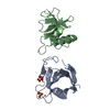 1rggS S: Starting model for refinement |
|---|---|
| Similar structure data |


-Links
 PDBj
PDBj- Assembly
| Deposited unit | 
| ||||||||
|---|---|---|---|---|---|---|---|---|---|
| 1 |
| ||||||||
| Unit cell |
|
-Components
| #1: Protein | Mass: 10555.467 Da / Num. of mol.: 1 / Mutation: N39S Source method: isolated from a genetically manipulated source Source: (gene. exp.) Streptomyces aureofaciens (bacteria) / Strain: BMK / Gene: U39467 / Plasmid: PUC19 / Production host: |
|---|---|
| #2: Water | ChemComp-HOH /  Mass: 18.015 Da / Num. of mol.: 95 / Source method: isolated from a natural source / Formula: H2O Mass: 18.015 Da / Num. of mol.: 95 / Source method: isolated from a natural source / Formula: H2O |
| Has protein modification | Y |
-Experimental details
-Experiment
| Experiment | Method: X-RAY DIFFRACTION / Number of used crystals: 1 |
|---|
- Sample preparation
| Crystal | Density Matthews: 2.37 Å3/Da / Density % sol: 48 % | |||||||||||||||||||||||||
|---|---|---|---|---|---|---|---|---|---|---|---|---|---|---|---|---|---|---|---|---|---|---|---|---|---|---|
| Crystal grow | pH: 8 Details: HANGING DROP VAPOUR EQUILIBRATION DROP: 10 MG/ML OF PROTEIN 0.1 M TRIS-HCL BUFFER AT PH 8.0 12,5 % PEG 6000 RESERVOIR: 25 % PEG 6000 | |||||||||||||||||||||||||
| Crystal grow | *PLUS Method: vapor diffusion, hanging drop | |||||||||||||||||||||||||
| Components of the solutions | *PLUS
|
-Data collection
| Diffraction | Mean temperature: 293 K |
|---|---|
| Diffraction source | Source: ROTATING ANODE / Type: RIGAKU RU200 / Wavelength: 1.5418 |
| Detector | Type: MARRESEARCH / Detector: IMAGE PLATE / Date: Jul 15, 1997 / Details: MSC "LONG" MIRRORS |
| Radiation | Protocol: SINGLE WAVELENGTH / Monochromatic (M) / Laue (L): M / Scattering type: x-ray |
| Radiation wavelength | Wavelength: 1.5418 Å / Relative weight: 1 |
| Reflection | Resolution: 1.6→14.9 Å / Num. obs: 12724 / % possible obs: 92.1 % / Redundancy: 4.8 % / Biso Wilson estimate: 25 Å2 / Rmerge(I) obs: 0.058 / Net I/σ(I): 30.1 |
| Reflection shell | Resolution: 1.6→1.63 Å / Redundancy: 2.6 % / Rmerge(I) obs: 0.471 / Mean I/σ(I) obs: 1.9 / % possible all: 57.1 |
| Reflection shell | *PLUS % possible obs: 57.1 % |
- Processing
| Software |
| ||||||||||||||||||||||||||||||||||||||||||||||||||||||||||||||||||||||||||||||||||||
|---|---|---|---|---|---|---|---|---|---|---|---|---|---|---|---|---|---|---|---|---|---|---|---|---|---|---|---|---|---|---|---|---|---|---|---|---|---|---|---|---|---|---|---|---|---|---|---|---|---|---|---|---|---|---|---|---|---|---|---|---|---|---|---|---|---|---|---|---|---|---|---|---|---|---|---|---|---|---|---|---|---|---|---|---|---|
| Refinement | Method to determine structure: MOLECULAR REPLACEMENT Starting model: PDB ENTRY 1RGG Resolution: 1.6→14.9 Å / Cross valid method: THROUGHOUT / σ(F): 0 / ESU R: 0.084
| ||||||||||||||||||||||||||||||||||||||||||||||||||||||||||||||||||||||||||||||||||||
| Displacement parameters | Biso mean: 26.2 Å2 | ||||||||||||||||||||||||||||||||||||||||||||||||||||||||||||||||||||||||||||||||||||
| Refinement step | Cycle: LAST / Resolution: 1.6→14.9 Å
| ||||||||||||||||||||||||||||||||||||||||||||||||||||||||||||||||||||||||||||||||||||
| Refine LS restraints |
| ||||||||||||||||||||||||||||||||||||||||||||||||||||||||||||||||||||||||||||||||||||
| Software | *PLUS Name: REFMAC / Classification: refinement | ||||||||||||||||||||||||||||||||||||||||||||||||||||||||||||||||||||||||||||||||||||
| Refinement | *PLUS Num. reflection obs: 11829 | ||||||||||||||||||||||||||||||||||||||||||||||||||||||||||||||||||||||||||||||||||||
| Solvent computation | *PLUS | ||||||||||||||||||||||||||||||||||||||||||||||||||||||||||||||||||||||||||||||||||||
| Displacement parameters | *PLUS |
