 Movie
Movie Controller

[English] 日本語
 Yorodumi
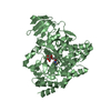
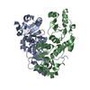
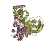
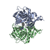
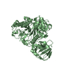
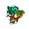
- PDB-1h9g: FadR, FATTY ACID RESPONSIVE TRANSCRIPTION FACTOR FROM E. COLI, in... -
+ Open data

- Basic information
| Entry | Database: PDB / ID: 1h9g | ||||||
|---|---|---|---|---|---|---|---|
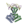
| Title | FadR, FATTY ACID RESPONSIVE TRANSCRIPTION FACTOR FROM E. COLI, in complex with myristoyl-CoA | ||||||
 Components Components | FATTY ACID METABOLISM REGULATOR PROTEIN | ||||||
 Keywords Keywords | TRANSCRIPTIONAL REGULATION | ||||||
| Function / homology |  Function and homology information Function and homology informationfatty-acyl-CoA binding / regulation of fatty acid metabolic process / positive regulation of fatty acid biosynthetic process / cis-regulatory region sequence-specific DNA binding / fatty acid metabolic process / DNA-binding transcription factor activity / negative regulation of DNA-templated transcription / regulation of DNA-templated transcription / positive regulation of DNA-templated transcription / protein homodimerization activity ...fatty-acyl-CoA binding / regulation of fatty acid metabolic process / positive regulation of fatty acid biosynthetic process / cis-regulatory region sequence-specific DNA binding / fatty acid metabolic process / DNA-binding transcription factor activity / negative regulation of DNA-templated transcription / regulation of DNA-templated transcription / positive regulation of DNA-templated transcription / protein homodimerization activity / DNA binding / cytosol Similarity search - Function | ||||||
| Biological species |  | ||||||
| Method |  X-RAY DIFFRACTION / SYNCHROTRON / MOLECULAR REPLACEMENT / Resolution: 2.1 Å X-RAY DIFFRACTION / SYNCHROTRON / MOLECULAR REPLACEMENT / Resolution: 2.1 Å | ||||||
 Authors Authors | Van Aalten, D.M.F. / Dirusso, C.C. / Knudsen, J. | ||||||
 Citation Citation | Journal: Embo J. / Year: 2001 Title: The Structural Basis of Acyl Coenzyme A-Dependent Regulation of the Transcription Factor Fadr Authors: Van Aalten, D.M.F. / Dirusso, C.C. / Knudsen, J. #1: Journal: Embo J. / Year: 2000Title: Crystal Structure of Fadr, a Fatty Acid-Responsive Transcription Factor with a Novel Acyl Coenzyme A-Binding Fold Authors: Van Aalten, D.M. / Dirusso, C.C. / Knudsen, J. / Wierenga, R.K. #2: Journal: Acta Crystallogr.,Sect.D / Year: 2000 Title: Crystallization and X-Ray Diffraction Studies of the Fatty-Acid Responsive Transcription Factor Fadr from Escherichia Coli Authors: Van Aalten, D.M.F. / Knudsen, J. / Dirusso, C.C. / Kokko, T. / Wierenga, R.K. | ||||||
| History |
|
- Structure visualization
| Structure viewer | Molecule: MolmilJmol/JSmol |
|---|
- Downloads & links
-Download
| PDBx/mmCIF format | 1h9g.cif.gz | 64.9 KB | Display | PDBx/mmCIF format |
|---|---|---|---|---|
| PDB format | pdb1h9g.ent.gz | 46.4 KB | Display | PDB format |
| PDBx/mmJSON format | 1h9g.json.gz | Tree view | PDBx/mmJSON format | |
| Others |  Other downloads Other downloads |
-Validation report
| Arichive directory | https://data.pdbj.org/pub/pdb/validation_reports/h9/1h9gftp://data.pdbj.org/pub/pdb/validation_reports/h9/1h9g | HTTPS FTP |
|---|
-Related structure data
| Related structure data | 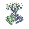 1h9tC  1e2xS S: Starting model for refinement C: citing same article ( |
|---|---|
| Similar structure data |


-Links
 PDBj
PDBj


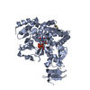
- Assembly
| Deposited unit | 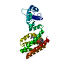
| ||||||||
|---|---|---|---|---|---|---|---|---|---|
| 1 | 
| ||||||||
| Unit cell |
| ||||||||
| Components on special symmetry positions |
|
-Components
| #1: Protein | Mass: 27493.111 Da / Num. of mol.: 1 / Fragment: FULL LENGTH Source method: isolated from a genetically manipulated source Source: (gene. exp.) |
|---|---|
| #2: Chemical | ChemComp-COA /   Mass: 767.534 Da / Num. of mol.: 1 / Source method: obtained synthetically / Formula: C21H36N7O16P3S Mass: 767.534 Da / Num. of mol.: 1 / Source method: obtained synthetically / Formula: C21H36N7O16P3S |
| #3: Chemical | ChemComp-MYR /   Mass: 228.371 Da / Num. of mol.: 1 / Source method: obtained synthetically / Formula: C14H28O2 Mass: 228.371 Da / Num. of mol.: 1 / Source method: obtained synthetically / Formula: C14H28O2 |
| #4: Water | ChemComp-HOH /  Mass: 18.015 Da / Num. of mol.: 134 / Source method: isolated from a natural source / Formula: H2O Mass: 18.015 Da / Num. of mol.: 134 / Source method: isolated from a natural source / Formula: H2O |
-Experimental details
-Experiment
| Experiment | Method: X-RAY DIFFRACTION / Number of used crystals: 1 |
|---|
- Sample preparation
| Crystal | Density Matthews: 2.7 Å3/Da / Density % sol: 54.4 % | |||||||||||||||||||||||||||||||||||||||||||||||||
|---|---|---|---|---|---|---|---|---|---|---|---|---|---|---|---|---|---|---|---|---|---|---|---|---|---|---|---|---|---|---|---|---|---|---|---|---|---|---|---|---|---|---|---|---|---|---|---|---|---|---|
| Crystal grow | pH: 7 / Details: pH 7.00 | |||||||||||||||||||||||||||||||||||||||||||||||||
| Crystal grow | *PLUS Temperature: 20 ℃ / pH: 8 / Method: vapor diffusion, hanging drop | |||||||||||||||||||||||||||||||||||||||||||||||||
| Components of the solutions | *PLUS
|
-Data collection
| Diffraction | Mean temperature: 100 K |
|---|---|
| Diffraction source | Source: SYNCHROTRON / Site: EMBL/DESY, HAMBURG  / Beamline: X11 / Wavelength: 0.93 / Beamline: X11 / Wavelength: 0.93 |
| Detector | Type: MARRESEARCH / Detector: CCD / Date: Aug 2, 2000 |
| Radiation | Protocol: SINGLE WAVELENGTH / Monochromatic (M) / Laue (L): M / Scattering type: x-ray |
| Radiation wavelength | Wavelength: 0.93 Å / Relative weight: 1 |
| Reflection | Resolution: 2.1→30 Å / Num. obs: 18556 / % possible obs: 97.8 % / Redundancy: 4.7 % / Biso Wilson estimate: 31.9 Å2 / Rmerge(I) obs: 0.038 / Net I/σ(I): 17.9 |
| Reflection shell | Resolution: 2.1→2.18 Å / Rmerge(I) obs: 0.292 / Mean I/σ(I) obs: 5 / % possible all: 95.5 |
| Reflection shell | *PLUS % possible obs: 95.5 % |
- Processing
| Software |
| ||||||||||||||||||||||||||||||||||||||||||||||||||||||||||||||||||||||||||||||||
|---|---|---|---|---|---|---|---|---|---|---|---|---|---|---|---|---|---|---|---|---|---|---|---|---|---|---|---|---|---|---|---|---|---|---|---|---|---|---|---|---|---|---|---|---|---|---|---|---|---|---|---|---|---|---|---|---|---|---|---|---|---|---|---|---|---|---|---|---|---|---|---|---|---|---|---|---|---|---|---|---|---|
| Refinement | Method to determine structure: MOLECULAR REPLACEMENT Starting model: 1E2X Resolution: 2.1→29.68 Å / Rfactor Rfree error: 0.01 / Data cutoff high absF: 2029989.25 / Isotropic thermal model: RESTRAINED / Cross valid method: THROUGHOUT / σ(F): 0 Details: SOME RESIDUES TRUNCATED TO ALA O DUE TO POORLY DEFINED ELECTRON DENSITY
| ||||||||||||||||||||||||||||||||||||||||||||||||||||||||||||||||||||||||||||||||
| Solvent computation | Solvent model: FLAT MODEL / Bsol: 82 Å2 / ksol: 0.431178 e/Å3 | ||||||||||||||||||||||||||||||||||||||||||||||||||||||||||||||||||||||||||||||||
| Displacement parameters | Biso mean: 43.6 Å2
| ||||||||||||||||||||||||||||||||||||||||||||||||||||||||||||||||||||||||||||||||
| Refine analyze |
| ||||||||||||||||||||||||||||||||||||||||||||||||||||||||||||||||||||||||||||||||
| Refinement step | Cycle: LAST / Resolution: 2.1→29.68 Å
| ||||||||||||||||||||||||||||||||||||||||||||||||||||||||||||||||||||||||||||||||
| Refine LS restraints |
| ||||||||||||||||||||||||||||||||||||||||||||||||||||||||||||||||||||||||||||||||
| LS refinement shell | Resolution: 2.1→2.23 Å / Rfactor Rfree error: 0.032 / Total num. of bins used: 6
| ||||||||||||||||||||||||||||||||||||||||||||||||||||||||||||||||||||||||||||||||
| Xplor file |
| ||||||||||||||||||||||||||||||||||||||||||||||||||||||||||||||||||||||||||||||||
| Software | *PLUS Name: CNS / Version: 1 / Classification: refinement | ||||||||||||||||||||||||||||||||||||||||||||||||||||||||||||||||||||||||||||||||
| Refine LS restraints | *PLUS
|
