[English] 日本語
Yorodumi
- PDB-2icy: Crystal Structure of a Putative UDP-glucose Pyrophosphorylase fro... -

+
Open data


ID or keywords:

Loading...

-
Basic information

Entry
Database: PDB / ID: 2icy
TitleCrystal Structure of a Putative UDP-glucose Pyrophosphorylase from Arabidopsis Thaliana with Bound UDP-glucose
ComponentsProbable UTP-glucose-1-phosphate uridylyltransferase 2
KeywordsTRANSFERASE / At3g03250 / UDP / putative UDP-glucose pyrophosphorylase / Structural Genomics Functional Follow-up Study / Structural Genomics / Protein Structure Initiative / PSI / CESG / Center for Eukaryotic Structural Genomics
Function / homology
Function and homology information


callose deposition in cell wall / pollen tube / sucrose metabolic process / UTP-glucose-1-phosphate uridylyltransferase / UTP:glucose-1-phosphate uridylyltransferase activity / pollen development / UDP-glucose metabolic process / cellular response to phosphate starvation / glycogen metabolic process / plasma membrane / cytosol
Similarity search - Function
UTP--glucose-1-phosphate uridylyltransferase / UDPGP family / UTP--glucose-1-phosphate uridylyltransferase / Hexapeptide repeat proteins / UDP N-Acetylglucosamine Acyltransferase; domain 1 / 3 Solenoid / Spore Coat Polysaccharide Biosynthesis Protein SpsA; Chain A / Spore Coat Polysaccharide Biosynthesis Protein SpsA; Chain A / Nucleotide-diphospho-sugar transferases / Alpha-Beta Complex ...UTP--glucose-1-phosphate uridylyltransferase / UDPGP family / UTP--glucose-1-phosphate uridylyltransferase / Hexapeptide repeat proteins / UDP N-Acetylglucosamine Acyltransferase; domain 1 / 3 Solenoid / Spore Coat Polysaccharide Biosynthesis Protein SpsA; Chain A / Spore Coat Polysaccharide Biosynthesis Protein SpsA; Chain A / Nucleotide-diphospho-sugar transferases / Alpha-Beta Complex / Mainly Beta / Alpha Beta
Similarity search - Domain/homology
URIDINE-5'-MONOPHOSPHATE / URIDINE-5'-DIPHOSPHATE-GLUCOSE / UTP--glucose-1-phosphate uridylyltransferase 2
Similarity search - Component
Biological speciesArabidopsis thaliana (thale cress)
MethodX-RAY DIFFRACTION / SYNCHROTRON / MOLECULAR REPLACEMENT / Resolution: 1.64 Å
AuthorsMcCoy, J.G. / Wesenberg, G.E. / Phillips Jr., G.N. / Bitto, E. / Bingman, C.A. / Center for Eukaryotic Structural Genomics (CESG)
CitationJournal: J.Mol.Biol. / Year: 2007
Title: Structure and Dynamics of UDP-Glucose Pyrophosphorylase from Arabidopsis thaliana with Bound UDP-Glucose and UTP.
Authors: McCoy, J.G. / Bitto, E. / Bingman, C.A. / Wesenberg, G.E. / Bannen, R.M. / Kondrashov, D.A. / Phillips Jr., G.N.
History
DepositionSep 13, 2006Deposition site: RCSB / Processing site: RCSB
Revision 1.0Oct 3, 2006Provider: repository / Type: Initial release
Revision 1.1Apr 1, 2008Group: Version format compliance
Revision 1.2Jul 13, 2011Group: Version format compliance
Revision 1.3Sep 9, 2015Group: Version format compliance
Revision 1.4Aug 30, 2023Group: Data collection / Database references ...Data collection / Database references / Derived calculations / Refinement description
Category: chem_comp_atom / chem_comp_bond ...chem_comp_atom / chem_comp_bond / database_2 / pdbx_initial_refinement_model / software / struct_site
Item: _database_2.pdbx_DOI / _database_2.pdbx_database_accession ..._database_2.pdbx_DOI / _database_2.pdbx_database_accession / _software.classification / _software.contact_author / _software.contact_author_email / _software.date / _software.language / _software.location / _software.name / _software.type / _software.version / _struct_site.pdbx_auth_asym_id / _struct_site.pdbx_auth_comp_id / _struct_site.pdbx_auth_seq_id

-
Structure visualization

Structure viewerMolecule:
MolmilJmol/JSmol

Downloads & links

-
Assembly

Deposited unit
A: Probable UTP-glucose-1-phosphate uridylyltransferase 2
B: Probable UTP-glucose-1-phosphate uridylyltransferase 2
hetero molecules


Theoretical massNumber of molelcules
Total (without water)105,0436
Polymers103,5082
Non-polymers1,5354
Water15,277848
1
A: Probable UTP-glucose-1-phosphate uridylyltransferase 2
hetero molecules


Theoretical massNumber of molelcules
Total (without water)52,3993
Polymers51,7541
Non-polymers6442
Water181
TypeNameSymmetry operationNumber
identity operation1_555x,y,z1
2
B: Probable UTP-glucose-1-phosphate uridylyltransferase 2
hetero molecules


Theoretical massNumber of molelcules
Total (without water)52,3202
Polymers51,7541
Non-polymers5661
Water181
TypeNameSymmetry operationNumber
identity operation1_555x,y,z1
Unit cell
Length a, b, c (Å)188.008, 59.712, 89.762
Angle α, β, γ (deg.)90.000, 100.32, 90.000
Int Tables number5
Space group name H-MC121
Number of models2
Components on special symmetry positions
IDModelComponents
11A-1226-

HOH

22A-1226-

HOH

-
Components

#1: Protein Probable UTP-glucose-1-phosphate uridylyltransferase 2 / UDP-glucose pyrophosphorylase 2 / UDPGP 2 / UGPase 2


Mass: 51754.090 Da / Num. of mol.: 2
Source method: isolated from a genetically manipulated source
Source: (gene. exp.) Arabidopsis thaliana (thale cress) / Gene: At3g03250, T17B22.6 / Plasmid: PVP-17 / Production host: Escherichia coli (E. coli) / Strain (production host): B834 P(RARE2)
References: UniProt: Q9M9P3, UTP-glucose-1-phosphate uridylyltransferase
#2: Chemical ChemComp-DMS / DIMETHYL SULFOXIDE


Mass: 78.133 Da / Num. of mol.: 1 / Source method: obtained synthetically / Formula: C2H6OS / Comment: DMSO, precipitant*YM
#3: Chemical ChemComp-UPG / URIDINE-5'-DIPHOSPHATE-GLUCOSE / URIDINE-5'-MONOPHOSPHATE GLUCOPYRANOSYL-MONOPHOSPHATE ESTER


Mass: 566.302 Da / Num. of mol.: 2 / Source method: obtained synthetically / Formula: C15H24N2O17P2
#4: Chemical ChemComp-U5P / URIDINE-5'-MONOPHOSPHATE


Mass: 324.181 Da / Num. of mol.: 1 / Source method: obtained synthetically / Formula: C9H13N2O9P
#5: Water ChemComp-HOH / water


Mass: 18.015 Da / Num. of mol.: 848 / Source method: isolated from a natural source / Formula: H2O

-
Experimental details

-
Experiment

ExperimentMethod: X-RAY DIFFRACTION / Number of used crystals: 1

-
Sample preparation

CrystalDensity Matthews: 2.39 Å3/Da / Density % sol: 48.6 %
Crystal growTemperature: 273 K / Method: vapor diffusion, hanging drop with micro-seeding
Details: Protein solution (10 mg/ml protein, 0.050 M sodium chloride, 0.0003 M TCEP, 0.005 M Tris PH 8.0) mixed in a 1:1 ratio with Well solution ( 28 % PEG 2K, 5% DMSO, 0.10 M MES/Acetate pH 5.5), ...Details: Protein solution (10 mg/ml protein, 0.050 M sodium chloride, 0.0003 M TCEP, 0.005 M Tris PH 8.0) mixed in a 1:1 ratio with Well solution ( 28 % PEG 2K, 5% DMSO, 0.10 M MES/Acetate pH 5.5), crystals soaked in well solution supplemented with 0.002 M UDP-glucose, vapor diffusion, hanging drop with micro-seeding, temperature 273K

-
Data collection

DiffractionMean temperature: 100 K
Diffraction sourceSource: SYNCHROTRON / Site: APS / Beamline: 22-ID / Wavelength: 0.97911 Å
DetectorType: MARMOSAIC 300 mm CCD / Detector: CCD / Date: Oct 11, 2005
Details: HORIZONTAL SAGITALLY FOCUSING 2ND BENT MONOCHROMATOR CRYSTAL, VERTICAL BENT FOCUSING MIRROR
RadiationMonochromator: CRYOGENICALLY COOLED SI (220) DOUBLE BOUNCE / Protocol: SINGLE WAVELENGTH / Monochromatic (M) / Laue (L): M / Scattering type: x-ray
Radiation wavelengthWavelength: 0.97911 Å / Relative weight: 1
ReflectionResolution: 1.64→38.25 Å / Num. all: 118994 / Num. obs: 118994 / % possible obs: 98.8 % / Observed criterion σ(F): 0 / Redundancy: 4.3 % / Biso Wilson estimate: 23 Å2 / Limit h max: 112 / Limit h min: -114 / Limit k max: 36 / Limit k min: -114 / Limit l max: 54 / Limit l min: 0 / Observed criterion F max: 2130663.21 / Observed criterion F min: 9.7 / Rmerge(I) obs: 0.042 / Χ2: 1.004 / Net I/σ(I): 20.7
Reflection shell

Diffraction-ID: 1

Resolution (Å)Redundancy (%)Rmerge(I) obsMean I/σ(I) obsNum. unique allΧ2% possible all
1.64-1.683.30.2953.89465940.98683.1
1.68-1.7240.25879461.00999.6
1.72-1.774.30.20980291.049100
1.77-1.824.30.17179681.041100
1.82-1.884.40.13280091.077100
1.88-1.944.40.10379611.061100
1.94-2.024.40.08279971.005100
2.02-2.114.40.07180081.036100
2.11-2.234.40.06680091.006100
2.23-2.374.40.05880410.967100
2.37-2.554.50.04780220.992100
2.55-2.84.50.04280470.957100
2.8-3.214.50.04480780.949100
3.21-4.044.40.03380981.086100
4.04-504.20.03281870.82499

-
Phasing

Phasing MRRfactor: 0.54 / Cor.coef. Fo:Fc: 0.229
Highest resolutionLowest resolution
Rotation3.5 Å46.61 Å
Translation3.5 Å46.61 Å

-
Processing

Software
NameVersionClassificationNB
DENZOdata reduction
SCALEPACKdata scaling
MOLREPphasing
CNS1.1refinement
PDB_EXTRACT2data extraction
HKL-2000data reduction
RefinementMethod to determine structure: MOLECULAR REPLACEMENT
Starting model: pdb entry 1Z90
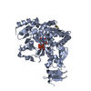
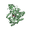
Resolution: 1.64→38.25 Å / Rfactor Rfree error: 0.003 / Occupancy max: 1 / Occupancy min: 0.3 / Cross valid method: THROUGHOUT
Details: The structure was refined using multiple conformers. Chains A and B of MODEL 1 and Chains A and B of MODEL 2 were refined as alternate conformers such that they both contributed to the total ...Details: The structure was refined using multiple conformers. Chains A and B of MODEL 1 and Chains A and B of MODEL 2 were refined as alternate conformers such that they both contributed to the total structure factor but did not interact with each other. The multiple models were necessary to account for the observable electron density and shows conformational changes that occur during the binding of UDP-glucose, with UDP-glucose bound (UPG 902) in chain B of model 1 and uridine-5'-monophosphate (U5G 903) in chain B of model 2. Waters common to both models are listed under both MODEL 1 and MODEL 2 with partial occupancies of 0.5. waters unique to MODEL 1 are assigned residue numbers 1001 TO 1050 and waters unique to MODEL 2 are assigned residue numbers 2001 TO 2006.
RfactorNum. reflection% reflectionSelection details
Rfree0.223 5964 5 %random
Rwork0.183 ---
all-119685 --
obs-118994 99.4 %-
Solvent computationSolvent model: CNS bulk solvent model used / Bsol: 44.3135 Å2 / ksol: 0.305117 e/Å3
Displacement parametersBiso max: 77.55 Å2 / Biso mean: 25.74 Å2 / Biso min: 5.34 Å2
Baniso -1Baniso -2Baniso -3
1--1.04 Å20 Å2-0.6 Å2
2--2.32 Å20 Å2
3----1.27 Å2
Refine analyze
FreeObs
Luzzati coordinate error0.21 Å0.16 Å
Luzzati d res low-5 Å
Luzzati sigma a0.17 Å0.1 Å
Luzzati d res high-1.64
Refinement stepCycle: LAST / Resolution: 1.64→38.25 Å
ProteinNucleic acidLigandSolventTotal
Num. atoms7152 0 76 848 8076
Refine LS restraints
Refine-IDTypeDev ideal
X-RAY DIFFRACTIONc_bond_d0.019
X-RAY DIFFRACTIONc_angle_deg2
X-RAY DIFFRACTIONc_dihedral_angle_d24.8
X-RAY DIFFRACTIONc_improper_angle_d1.3
LS refinement shell

Refine-ID: X-RAY DIFFRACTION / Total num. of bins used: 8

Resolution (Å)Rfactor RfreeNum. reflection Rfree% reflection Rfree (%)Rfactor RworkNum. reflection RworkRfactor Rfree errorNum. reflection allNum. reflection obs% reflection obs (%)
1.64-1.720.3197375.20.266134880.012148781422595.6
1.72-1.810.29174550.214141550.011149381490099.7
1.81-1.920.26974950.192141420.01149261489199.8
1.92-2.070.247855.30.187140950.009148971488099.9
2.07-2.280.22274550.179141860.008149401493199.9
2.28-2.610.2357214.80.176142410.009149711496299.9
2.61-3.280.2157144.80.181143150.0081503415029100
3.28-38.250.1997685.10.174144080.007152521517699.5

+
About Yorodumi

-
News

-
Feb 9, 2022. New format data for meta-information of EMDB entries

New format data for meta-information of EMDB entries

  • Version 3 of the EMDB header file is now the official format.
  • The previous official version 1.9 will be removed from the archive.

Related info.:EMDB header

External links:wwPDB to switch to version 3 of the EMDB data model

-
Aug 12, 2020. Covid-19 info

Covid-19 info

URL: https://pdbjlvh1.pdbj.org/emnavi/covid19.php

New page: Covid-19 featured information page in EM Navigator.

Related info.:Covid-19 info / Mar 5, 2020. Novel coronavirus structure data

+
Mar 5, 2020. Novel coronavirus structure data

Novel coronavirus structure data

Related info.:Yorodumi Speices / Aug 12, 2020. Covid-19 info

External links:COVID-19 featured content - PDBj / Molecule of the Month (242):Coronavirus Proteases

+
Jan 31, 2019. EMDB accession codes are about to change! (news from PDBe EMDB page)

EMDB accession codes are about to change! (news from PDBe EMDB page)

  • The allocation of 4 digits for EMDB accession codes will soon come to an end. Whilst these codes will remain in use, new EMDB accession codes will include an additional digit and will expand incrementally as the available range of codes is exhausted. The current 4-digit format prefixed with “EMD-” (i.e. EMD-XXXX) will advance to a 5-digit format (i.e. EMD-XXXXX), and so on. It is currently estimated that the 4-digit codes will be depleted around Spring 2019, at which point the 5-digit format will come into force.
  • The EM Navigator/Yorodumi systems omit the EMD- prefix.

Related info.:Q: What is EMD? / ID/Accession-code notation in Yorodumi/EM Navigator

External links:EMDB Accession Codes are Changing Soon! / Contact to PDBj

+
Jul 12, 2017. Major update of PDB

Major update of PDB

  • wwPDB released updated PDB data conforming to the new PDBx/mmCIF dictionary.
  • This is a major update changing the version number from 4 to 5, and with Remediation, in which all the entries are updated.
  • In this update, many items about electron microscopy experimental information are reorganized (e.g. em_software).
  • Now, EM Navigator and Yorodumi are based on the updated data.

External links:wwPDB Remediation / Enriched Model Files Conforming to OneDep Data Standards Now Available in the PDB FTP Archive

-
Yorodumi

Thousand views of thousand structures

  • Yorodumi is a browser for structure data from EMDB, PDB, SASBDB, etc.
  • This page is also the successor to EM Navigator detail page, and also detail information page/front-end page for Omokage search.
  • The word "yorodu" (or yorozu) is an old Japanese word meaning "ten thousand". "mi" (miru) is to see.

Related info.:EMDB / PDB / SASBDB / Comparison of 3 databanks / Yorodumi Search / Aug 31, 2016. New EM Navigator & Yorodumi / Yorodumi Papers / Jmol/JSmol / Function and homology information / Changes in new EM Navigator and Yorodumi

Read more