- PDB-1e2x: FadR, fatty acid responsive transcription factor from E. coli -
+
Open data
ID or keywords:
Loading...
-
Basic information
Entry
Database: PDB / ID: 1e2x
Title
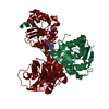
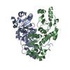
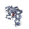
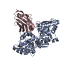
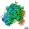
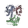
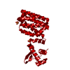
FadR, fatty acid responsive transcription factor from E. coli
Components
FATTY ACID METABOLISM REGULATOR PROTEIN
Keywords
TRANSCRIPTIONAL REGULATION
Function / homology
Function and homology information
fatty-acyl-CoA binding / regulation of fatty acid metabolic process / positive regulation of fatty acid biosynthetic process / cis-regulatory region sequence-specific DNA binding / fatty acid metabolic process / DNA-binding transcription factor activity / negative regulation of DNA-templated transcription / regulation of DNA-templated transcription / positive regulation of DNA-templated transcription / protein homodimerization activity ...fatty-acyl-CoA binding / regulation of fatty acid metabolic process / positive regulation of fatty acid biosynthetic process / cis-regulatory region sequence-specific DNA binding / fatty acid metabolic process / DNA-binding transcription factor activity / negative regulation of DNA-templated transcription / regulation of DNA-templated transcription / positive regulation of DNA-templated transcription / protein homodimerization activity / DNA binding / cytosol Similarity search - Function
Mass: 27493.111 Da / Num. of mol.: 1 / Fragment: FULL LENGTH Source method: isolated from a genetically manipulated source Source: (gene. exp.) ESCHERICHIA COLI (E. coli) / Production host: ESCHERICHIA COLI (E. coli) / References: UniProt: P09371, UniProt: P0A8V6*PLUS
Resolution: 2→25 Å / Num. obs: 18125 / % possible obs: 95.2 % / Redundancy: 3.2 % / Biso Wilson estimate: 16.9 Å2 / Rmerge(I) obs: 0.07 / Net I/σ(I): 9.7
Reflection shell
Resolution: 2→2.07 Å / Rmerge(I) obs: 0.448 / Mean I/σ(I) obs: 2.6 / % possible all: 74
Reflection shell
*PLUS
% possible obs: 73.8 %
-
Processing
Software
Name
Version
Classification
CNS
1
refinement
DENZO
datareduction
SCALEPACK
datascaling
CNS
1
phasing
Refinement
Method to determine structure: MAD / Resolution: 2→24.48 Å / Rfactor Rfree error: 0.009 / Data cutoff high absF: 1880425.42 / Isotropic thermal model: RESTRAINED / Cross valid method: THROUGHOUT / σ(F): 0 Details: SOME RESIDUES TRUNCATED TO ALA OR GLY DUE TO POORLY DEFINED ELECTRON DENSITY
In the structure databanks used in Yorodumi, some data are registered as the other names, "COVID-19 virus" and "2019-nCoV". Here are the details of the virus and the list of structure data.
Jan 31, 2019. EMDB accession codes are about to change! (news from PDBe EMDB page)
EMDB accession codes are about to change! (news from PDBe EMDB page)
The allocation of 4 digits for EMDB accession codes will soon come to an end. Whilst these codes will remain in use, new EMDB accession codes will include an additional digit and will expand incrementally as the available range of codes is exhausted. The current 4-digit format prefixed with “EMD-” (i.e. EMD-XXXX) will advance to a 5-digit format (i.e. EMD-XXXXX), and so on. It is currently estimated that the 4-digit codes will be depleted around Spring 2019, at which point the 5-digit format will come into force.
The EM Navigator/Yorodumi systems omit the EMD- prefix.
Related info.:Q: What is EMD? / ID/Accession-code notation in Yorodumi/EM Navigator
Yorodumi is a browser for structure data from EMDB, PDB, SASBDB, etc.
This page is also the successor to EM Navigator detail page, and also detail information page/front-end page for Omokage search.
The word "yorodu" (or yorozu) is an old Japanese word meaning "ten thousand". "mi" (miru) is to see.
Related info.:EMDB / PDB / SASBDB / Comparison of 3 databanks / Yorodumi Search / Aug 31, 2016. New EM Navigator & Yorodumi / Yorodumi Papers / Jmol/JSmol / Function and homology information / Changes in new EM Navigator and Yorodumi
 Movie
Movie Controller


 Open data
 Basic information
 Components
 Keywords
 Function and homology information
 X-RAY DIFFRACTION /
  Authors
 Citation
 Structure visualization
 Downloads & links
 Other downloads


 PDBj
PDBj Assembly
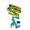

 Mass: 96.063 Da / Num. of mol.: 2 / Source method: obtained synthetically / Formula: SO4
 Mass: 18.015 Da / Num. of mol.: 156 / Source method: isolated from a natural source / Formula: H2O
 Sample preparation
 / Beamline: BM14 / Wavelength: 0.9535, 1.0
 Processing