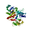
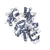
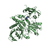
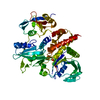
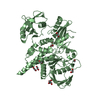
[English] 日本語
Yorodumi
- PDB-4dgp: The wild-type Src homology 2 (SH2)-domain containing protein tyro... -

+
Open data


ID or keywords:

Loading...

-
Basic information

Entry
Database: PDB / ID: 4dgp
TitleThe wild-type Src homology 2 (SH2)-domain containing protein tyrosine phosphatase-2 (SHP2)
ComponentsTyrosine-protein phosphatase non-receptor type 11
KeywordsHYDROLASE
Function / homology
Function and homology information


negative regulation of cortisol secretion / intestinal epithelial cell migration / microvillus organization / negative regulation of growth hormone secretion / genitalia development / atrioventricular canal development / negative regulation of cell adhesion mediated by integrin / STAT5 Activation / Netrin mediated repulsion signals / positive regulation of hormone secretion ...negative regulation of cortisol secretion / intestinal epithelial cell migration / microvillus organization / negative regulation of growth hormone secretion / genitalia development / atrioventricular canal development / negative regulation of cell adhesion mediated by integrin / STAT5 Activation / Netrin mediated repulsion signals / positive regulation of hormone secretion / cerebellar cortex formation / multicellular organismal reproductive process / regulation of protein export from nucleus / positive regulation of ossification / hormone metabolic process / Interleukin-37 signaling / Signaling by Leptin / negative regulation of chondrocyte differentiation / MET activates PTPN11 / Regulation of RUNX1 Expression and Activity / face morphogenesis / ERBB signaling pathway / Costimulation by the CD28 family / Signal regulatory protein family interactions / peptide hormone receptor binding / negative regulation of type I interferon production / platelet formation / megakaryocyte development / organ growth / triglyceride metabolic process / CTLA4 inhibitory signaling / Platelet sensitization by LDL / Interleukin-20 family signaling / PI-3K cascade:FGFR3 / Interleukin-6 signaling / STAT5 activation downstream of FLT3 ITD mutants / PI-3K cascade:FGFR2 / PI-3K cascade:FGFR4 / Prolactin receptor signaling / PI-3K cascade:FGFR1 / MAPK3 (ERK1) activation / MAPK1 (ERK2) activation / PECAM1 interactions / Bergmann glial cell differentiation / regulation of cell adhesion mediated by integrin / regulation of type I interferon-mediated signaling pathway / phosphoprotein phosphatase activity / inner ear development / neurotrophin TRK receptor signaling pathway / platelet-derived growth factor receptor signaling pathway / non-membrane spanning protein tyrosine phosphatase activity / RET signaling / peptidyl-tyrosine dephosphorylation / Interleukin-3, Interleukin-5 and GM-CSF signaling / Regulation of IFNA/IFNB signaling / PI3K Cascade / PD-1 signaling / regulation of protein-containing complex assembly / ephrin receptor signaling pathway / fibroblast growth factor receptor signaling pathway / GAB1 signalosome / Activated NTRK2 signals through FRS2 and FRS3 / negative regulation of insulin secretion / Regulation of IFNG signaling / positive regulation of insulin receptor signaling pathway / FRS-mediated FGFR3 signaling / Signaling by CSF3 (G-CSF) / Signaling by FLT3 ITD and TKD mutants / homeostasis of number of cells within a tissue / FRS-mediated FGFR2 signaling / FRS-mediated FGFR4 signaling / Tie2 Signaling / FRS-mediated FGFR1 signaling / cellular response to epidermal growth factor stimulus / cell adhesion molecule binding / GPVI-mediated activation cascade / T cell costimulation / FLT3 Signaling / positive regulation of interferon-beta production / hormone-mediated signaling pathway / phosphotyrosine residue binding / positive regulation of mitotic cell cycle / Downstream signal transduction / protein dephosphorylation / Activation of IRF3, IRF7 mediated by TBK1, IKKε (IKBKE) / protein-tyrosine-phosphatase / axonogenesis / protein tyrosine kinase binding / DNA damage checkpoint signaling / protein tyrosine phosphatase activity / integrin-mediated signaling pathway / positive regulation of glucose import / Negative regulation of FGFR3 signaling / Negative regulation of FGFR2 signaling / Negative regulation of FGFR4 signaling / Negative regulation of FGFR1 signaling / Spry regulation of FGF signaling / insulin receptor binding / epidermal growth factor receptor signaling pathway / multicellular organism growth
Similarity search - Function
Protein-tyrosine phosphatase, non-receptor type-6, -11 / SH2 domain / SHC Adaptor Protein / Protein tyrosine phosphatase superfamily / Protein-Tyrosine Phosphatase; Chain A / Protein tyrosine phosphatase, catalytic domain / PTP type protein phosphatase domain profile. / Protein-tyrosine phosphatase / Tyrosine-specific protein phosphatase, PTPase domain / Protein-tyrosine phosphatase, catalytic ...Protein-tyrosine phosphatase, non-receptor type-6, -11 / SH2 domain / SHC Adaptor Protein / Protein tyrosine phosphatase superfamily / Protein-Tyrosine Phosphatase; Chain A / Protein tyrosine phosphatase, catalytic domain / PTP type protein phosphatase domain profile. / Protein-tyrosine phosphatase / Tyrosine-specific protein phosphatase, PTPase domain / Protein-tyrosine phosphatase, catalytic / Protein tyrosine phosphatase, catalytic domain motif / Tyrosine specific protein phosphatases active site. / Protein-tyrosine phosphatase, active site / Tyrosine-specific protein phosphatases domain / Tyrosine specific protein phosphatases domain profile. / Protein-tyrosine phosphatase-like / SH2 domain / Src homology 2 (SH2) domain profile. / Src homology 2 domains / SH2 domain / SH2 domain superfamily / Alpha-Beta Complex / 2-Layer Sandwich / Alpha Beta
Similarity search - Domain/homology
Tyrosine-protein phosphatase non-receptor type 11
Similarity search - Component
Biological speciesHomo sapiens (human)
MethodX-RAY DIFFRACTION / SYNCHROTRON / MOLECULAR REPLACEMENT / Resolution: 2.3 Å
AuthorsYu, Z.H. / Xu, J. / Walls, C.D. / Chen, L. / Zhang, S. / Wu, L. / Wang, L.N. / Liu, S.J. / Zhang, Z.Y.
CitationJournal: J.Biol.Chem. / Year: 2013
Title: Structural and Mechanistic Insights into LEOPARD Syndrome-Associated SHP2 Mutations.
Authors: Yu, Z.H. / Xu, J. / Walls, C.D. / Chen, L. / Zhang, S. / Zhang, R. / Wu, L. / Wang, L. / Liu, S. / Zhang, Z.Y.
History
DepositionJan 26, 2012Deposition site: RCSB / Processing site: RCSB
Revision 1.0Mar 6, 2013Provider: repository / Type: Initial release
Revision 1.1Mar 20, 2013Group: Database references
Revision 1.2May 1, 2013Group: Database references
Revision 1.3Feb 28, 2024Group: Data collection / Database references
Category: chem_comp_atom / chem_comp_bond ...chem_comp_atom / chem_comp_bond / database_2 / struct_ref_seq_dif
Item: _database_2.pdbx_DOI / _database_2.pdbx_database_accession / _struct_ref_seq_dif.details

-
Structure visualization

Structure viewerMolecule:
MolmilJmol/JSmol

Downloads & links

-
Assembly

Deposited unit
A: Tyrosine-protein phosphatase non-receptor type 11


Theoretical massNumber of molelcules
Total (without water)61,7621
Polymers61,7621
Non-polymers00
Water2,702150
1


  • Idetical with deposited unit
  • defined by software
TypeNameSymmetry operationNumber
identity operation1_555x,y,z1
MethodPISA
Unit cell
Length a, b, c (Å)54.853, 221.102, 40.358
Angle α, β, γ (deg.)90.00, 90.00, 90.00
Int Tables number18
Space group name H-MP21212

-
Components

#1: Protein Tyrosine-protein phosphatase non-receptor type 11 / SRC homology 2 (SH2)-domain containing protein tyrosine phosphatase-2 / SHP2 / Protein-tyrosine ...SRC homology 2 (SH2)-domain containing protein tyrosine phosphatase-2 / SHP2 / Protein-tyrosine phosphatase 1D / PTP-1D / Protein-tyrosine phosphatase 2C / PTP-2C / SH-PTP2 / SHP-2 / SH-PTP3


Mass: 61761.641 Da / Num. of mol.: 1
Fragment: N-SH2, C-SH2, and PTP domains (UNP residues 1-532)
Source method: isolated from a genetically manipulated source
Source: (gene. exp.) Homo sapiens (human) / Gene: PTPN11, PTP2C, SHPTP2 / Plasmid: pET21A+ / Production host: Escherichia coli (E. coli) / Strain (production host): BL21(DE3) / References: UniProt: Q06124, protein-tyrosine-phosphatase
#2: Water ChemComp-HOH / water


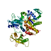
Mass: 18.015 Da / Num. of mol.: 150 / Source method: isolated from a natural source / Formula: H2O

-
Experimental details

-
Experiment

ExperimentMethod: X-RAY DIFFRACTION / Number of used crystals: 1

-
Sample preparation

CrystalDensity Matthews: 1.98 Å3/Da / Density % sol: 37.92 %
Crystal growTemperature: 293 K / Method: vapor diffusion, hanging drop / pH: 7.3
Details: 20% PEG3350, 300 mM potassium formate, pH 7.3, VAPOR DIFFUSION, HANGING DROP, temperature 293K

-
Data collection

DiffractionMean temperature: 100 K
Diffraction sourceSource: SYNCHROTRON / Site: APS / Beamline: 19-BM / Wavelength: 0.97915 Å
DetectorType: ADSC QUANTUM 210r / Detector: CCD
RadiationMonochromator: Si(111) / Protocol: SINGLE WAVELENGTH / Monochromatic (M) / Laue (L): M / Scattering type: x-ray
Radiation wavelengthWavelength: 0.97915 Å / Relative weight: 1
ReflectionResolution: 2.3→50 Å / Num. all: 22826 / Num. obs: 21799 / % possible obs: 95.6 % / Observed criterion σ(F): 0 / Observed criterion σ(I): 0 / Redundancy: 4.9 % / Rmerge(I) obs: 0.087 / Rsym value: 0.087 / Net I/σ(I): 25.28
Reflection shellResolution: 2.3→2.34 Å / Redundancy: 4.9 % / Rmerge(I) obs: 0.628 / Mean I/σ(I) obs: 2.9 / Rsym value: 0.628 / % possible all: 92.1

-
Processing

Software
NameVersionClassification
HKL-3000data collection
MOLREPphasing
PHENIX(phenix.refine: 1.6.4_486)refinement
HKL-3000data reduction
HKL-3000data scaling
RefinementMethod to determine structure: MOLECULAR REPLACEMENT / Resolution: 2.3→36.85 Å / SU ML: 0.32 / σ(F): 1.44 / Phase error: 25.7 / Stereochemistry target values: ML
RfactorNum. reflection% reflectionSelection details
Rfree0.2548 1085 4.99 %RANDOM
Rwork0.1962 ---
obs0.1991 21761 95.58 %-
all-21855 --
Solvent computationShrinkage radii: 0.83 Å / VDW probe radii: 1.1 Å / Solvent model: FLAT BULK SOLVENT MODEL / Bsol: 44.491 Å2 / ksol: 0.356 e/Å3
Displacement parameters
Baniso -1Baniso -2Baniso -3
1-7.2941 Å2-0 Å20 Å2
2---11.0538 Å2-0 Å2
3---3.7596 Å2
Refinement stepCycle: LAST / Resolution: 2.3→36.85 Å
ProteinNucleic acidLigandSolventTotal
Num. atoms4024 0 0 150 4174
Refine LS restraints
Refine-IDTypeDev idealNumber
X-RAY DIFFRACTIONf_bond_d0.0084106
X-RAY DIFFRACTIONf_angle_d1.1435537
X-RAY DIFFRACTIONf_chiral_restr0.074595
X-RAY DIFFRACTIONf_plane_restr0.004720
X-RAY DIFFRACTIONf_dihedral_angle_d16.0881544
LS refinement shell

Refine-ID: X-RAY DIFFRACTION

Resolution (Å)Rfactor RfreeNum. reflection RfreeRfactor RworkNum. reflection RworkNum. reflection obs% reflection obs (%)
2.3-2.40470.31691310.24762440257192
2.4047-2.53150.34491310.2512500263194
2.5315-2.690.33841300.25782538266896
2.69-2.89770.29691370.23592623276098
2.8977-3.18910.28661390.211426412780100
3.1891-3.65020.23861420.18232685282799
3.6502-4.59750.24181350.16252626276196
4.5975-36.8550.19811400.17962623276391

+
About Yorodumi

-
News

-
Feb 9, 2022. New format data for meta-information of EMDB entries

New format data for meta-information of EMDB entries

  • Version 3 of the EMDB header file is now the official format.
  • The previous official version 1.9 will be removed from the archive.

Related info.:EMDB header

External links:wwPDB to switch to version 3 of the EMDB data model

-
Aug 12, 2020. Covid-19 info

Covid-19 info

URL: https://pdbjlvh1.pdbj.org/emnavi/covid19.php

New page: Covid-19 featured information page in EM Navigator.

Related info.:Covid-19 info / Mar 5, 2020. Novel coronavirus structure data

+
Mar 5, 2020. Novel coronavirus structure data

Novel coronavirus structure data

Related info.:Yorodumi Speices / Aug 12, 2020. Covid-19 info

External links:COVID-19 featured content - PDBj / Molecule of the Month (242):Coronavirus Proteases

+
Jan 31, 2019. EMDB accession codes are about to change! (news from PDBe EMDB page)

EMDB accession codes are about to change! (news from PDBe EMDB page)

  • The allocation of 4 digits for EMDB accession codes will soon come to an end. Whilst these codes will remain in use, new EMDB accession codes will include an additional digit and will expand incrementally as the available range of codes is exhausted. The current 4-digit format prefixed with “EMD-” (i.e. EMD-XXXX) will advance to a 5-digit format (i.e. EMD-XXXXX), and so on. It is currently estimated that the 4-digit codes will be depleted around Spring 2019, at which point the 5-digit format will come into force.
  • The EM Navigator/Yorodumi systems omit the EMD- prefix.

Related info.:Q: What is EMD? / ID/Accession-code notation in Yorodumi/EM Navigator

External links:EMDB Accession Codes are Changing Soon! / Contact to PDBj

+
Jul 12, 2017. Major update of PDB

Major update of PDB

  • wwPDB released updated PDB data conforming to the new PDBx/mmCIF dictionary.
  • This is a major update changing the version number from 4 to 5, and with Remediation, in which all the entries are updated.
  • In this update, many items about electron microscopy experimental information are reorganized (e.g. em_software).
  • Now, EM Navigator and Yorodumi are based on the updated data.

External links:wwPDB Remediation / Enriched Model Files Conforming to OneDep Data Standards Now Available in the PDB FTP Archive

-
Yorodumi

Thousand views of thousand structures

  • Yorodumi is a browser for structure data from EMDB, PDB, SASBDB, etc.
  • This page is also the successor to EM Navigator detail page, and also detail information page/front-end page for Omokage search.
  • The word "yorodu" (or yorozu) is an old Japanese word meaning "ten thousand". "mi" (miru) is to see.

Related info.:EMDB / PDB / SASBDB / Comparison of 3 databanks / Yorodumi Search / Aug 31, 2016. New EM Navigator & Yorodumi / Yorodumi Papers / Jmol/JSmol / Function and homology information / Changes in new EM Navigator and Yorodumi

Read more