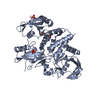
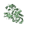
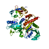
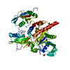
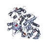
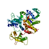
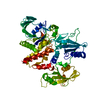
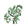
Entry Database : PDB / ID : 6atdTitle Oxidized SHP2 forms a disulfide bond between Cys367 and Cys459 Tyrosine-protein phosphatase non-receptor type 11 Keywords Function / homology Function Domain/homology Component
/ / / / / / / / / / / / / / / / / / / / / / / / / / / / / / / / / / / / / / / / / / / / / / / / / / / / / / / / / / / / / / / / / / / / / / / / / / / / / / / / / / / / / / / / / / / / / / / / / / / / / / / / / / / / / / / / / / / / / / / / / / / / / / / / / / / / / / / / / / / / Biological species Homo sapiens (human)Method / / / Resolution : 2.5 Å Authors Page, R. / Peti, W. / Critton, D.A. Funding support Organization Grant number Country American Cancer Society RSG-08-067-01-LIB
Journal : ACS Omega / Year : 2017Title : Redox Regulation of a Gain-of-Function Mutation (N308D) in SHP2 Noonan Syndrome.Authors : Machado, L.E.S.F. / Critton, D.A. / Page, R. / Peti, W. History Deposition Aug 28, 2017 Deposition site / Processing site Revision 1.0 Jul 11, 2018 Provider / Type Revision 1.1 Jan 29, 2020 Group / Category / Item Revision 1.2 Oct 4, 2023 Group / Database references / Refinement descriptionCategory chem_comp_atom / chem_comp_bond ... chem_comp_atom / chem_comp_bond / database_2 / pdbx_initial_refinement_model Item / _database_2.pdbx_database_accessionRevision 1.3 Nov 13, 2024 Group / Category / pdbx_modification_feature
Show all Show less
 Movie
Movie Controller


 Open data
 Basic information
 Components
 Keywords
 Function and homology information
 Homo sapiens (human)
 X-RAY DIFFRACTION /
  Authors
 United States, 1items
  Citation
 Structure visualization
 Downloads & links
 Other downloads
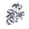


 PDBj
PDBj


 Assembly


 Mass: 94.971 Da / Num. of mol.: 4 / Source method: obtained synthetically / Formula: PO4
 Mass: 18.015 Da / Num. of mol.: 180 / Source method: isolated from a natural source / Formula: H2O
 Sample preparation
 Processing