 Movie
Movie Controller


+ Open data

- Basic information
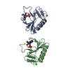
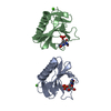
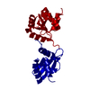
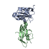
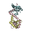
| Entry | Database: PDB / ID: 5x7b | ||||||
|---|---|---|---|---|---|---|---|
| Title | Crystal structure of SHP2_SH2-CagA EPIYA_C peptide complex | ||||||
 Components Components |
| ||||||
 Keywords Keywords | HYDROLASE / Helicobacter pylori CagA / SH2 domain-containing protein tyrosine phosphatase 2 (SHP2) / CagA polymorphism | ||||||
| Function / homology |  Function and homology information Function and homology informationsymbiont-mediated perturbation of host cell cycle progression / atrioventricular canal development / genitalia development / toxin transmembrane transporter activity / STAT5 Activation / Co-inhibition by BTLA / Netrin mediated repulsion signals / negative regulation of neutrophil activation / positive regulation of lipopolysaccharide-mediated signaling pathway / Interleukin-37 signaling ...symbiont-mediated perturbation of host cell cycle progression / atrioventricular canal development / genitalia development / toxin transmembrane transporter activity / STAT5 Activation / Co-inhibition by BTLA / Netrin mediated repulsion signals / negative regulation of neutrophil activation / positive regulation of lipopolysaccharide-mediated signaling pathway / Interleukin-37 signaling / Signaling by Leptin / negative regulation of cell adhesion mediated by integrin / positive regulation of ossification / MET activates PTPN11 / negative regulation of chondrocyte differentiation / face morphogenesis / Regulation of RUNX1 Expression and Activity / Signal regulatory protein family interactions / ERBB signaling pathway / Interleukin-20 family signaling / Interleukin-6 signaling / Co-inhibition by CTLA4 / PI-3K cascade:FGFR3 / STAT5 activation downstream of FLT3 ITD mutants / Platelet sensitization by LDL / negative regulation of T cell activation / PI-3K cascade:FGFR2 / peptide hormone receptor binding / PI-3K cascade:FGFR4 / MAPK3 (ERK1) activation / negative regulation of type I interferon production / PI-3K cascade:FGFR1 / regulation of type I interferon-mediated signaling pathway / MAPK1 (ERK2) activation / inner ear development / Prolactin receptor signaling / PECAM1 interactions / peptidyl-tyrosine dephosphorylation / non-membrane spanning protein tyrosine phosphatase activity / fibroblast growth factor receptor signaling pathway / Regulation of IFNA/IFNB signaling / RET signaling / positive regulation of intracellular signal transduction / Interleukin-3, Interleukin-5 and GM-CSF signaling / Co-inhibition by PD-1 / PI3K Cascade / ephrin receptor signaling pathway / positive regulation of insulin receptor signaling pathway / regulation of protein-containing complex assembly / Regulation of IFNG signaling / GAB1 signalosome / negative regulation of T cell receptor signaling pathway / negative regulation of T cell proliferation / Activated NTRK2 signals through FRS2 and FRS3 / GPVI-mediated activation cascade / T cell costimulation / Signaling by CSF3 (G-CSF) / FRS-mediated FGFR3 signaling / Signaling by FLT3 ITD and TKD mutants / FRS-mediated FGFR2 signaling / phosphotyrosine residue binding / FRS-mediated FGFR4 signaling / phosphoprotein phosphatase activity / FRS-mediated FGFR1 signaling / protein-tyrosine-phosphatase / Tie2 Signaling / FLT3 Signaling / protein tyrosine phosphatase activity / cell adhesion molecule binding / positive regulation of interferon-beta production / Downstream signal transduction / cellular response to epidermal growth factor stimulus / protein tyrosine kinase binding / positive regulation of D-glucose import across plasma membrane / insulin receptor binding / Activation of IRF3, IRF7 mediated by TBK1, IKKε (IKBKE) / Negative regulation of FGFR3 signaling / cellular response to mechanical stimulus / Negative regulation of FGFR2 signaling / Negative regulation of FGFR4 signaling / Negative regulation of FGFR1 signaling / brain development / Signaling by SCF-KIT / Spry regulation of FGF signaling / receptor tyrosine kinase binding / vasodilation / epidermal growth factor receptor signaling pathway / Constitutive Signaling by Aberrant PI3K in Cancer / cytokine-mediated signaling pathway / Signaling by CSF1 (M-CSF) in myeloid cells / Interferon alpha/beta signaling / positive regulation of tumor necrosis factor production / PIP3 activates AKT signaling / heart development / PI5P, PP2A and IER3 Regulate PI3K/AKT Signaling / signaling receptor complex adaptor activity / molecular adaptor activity / positive regulation of ERK1 and ERK2 cascade / positive regulation of phosphatidylinositol 3-kinase/protein kinase B signal transduction / cadherin binding Similarity search - Function | ||||||
| Biological species |  Homo sapiens (human) Homo sapiens (human)  Helicobacter pylori (bacteria) Helicobacter pylori (bacteria) | ||||||
| Method |  X-RAY DIFFRACTION / SYNCHROTRON / MOLECULAR REPLACEMENT / Resolution: 2.45 Å X-RAY DIFFRACTION / SYNCHROTRON / MOLECULAR REPLACEMENT / Resolution: 2.45 Å | ||||||
 Authors Authors | Senda, M. / Senda, T. | ||||||
 Citation Citation | Journal: Cell Rep / Year: 2017 Title: Differential Mechanisms for SHP2 Binding and Activation Are Exploited by Geographically Distinct Helicobacter pylori CagA Oncoproteins. Authors: Hayashi, T. / Senda, M. / Suzuki, N. / Nishikawa, H. / Ben, C. / Tang, C. / Nagase, L. / Inoue, K. / Senda, T. / Hatakeyama, M. | ||||||
| History |
|
- Structure visualization
| Structure viewer | Molecule: MolmilJmol/JSmol |
|---|
- Downloads & links
-Download
| PDBx/mmCIF format | 5x7b.cif.gz | 56.5 KB | Display | PDBx/mmCIF format |
|---|---|---|---|---|
| PDB format | pdb5x7b.ent.gz | 38.4 KB | Display | PDB format |
| PDBx/mmJSON format | 5x7b.json.gz | Tree view | PDBx/mmJSON format | |
| Others |  Other downloads Other downloads |
-Validation report
| Arichive directory | https://data.pdbj.org/pub/pdb/validation_reports/x7/5x7bftp://data.pdbj.org/pub/pdb/validation_reports/x7/5x7b | HTTPS FTP |
|---|
-Related structure data
| Related structure data |  5x94SC  5x7c S: Starting model for refinement C: citing same article ( |
|---|---|
| Similar structure data |


-Links
 PDBj
PDBj


- Assembly
| Deposited unit | 
| ||||||||
|---|---|---|---|---|---|---|---|---|---|
| 1 |
| ||||||||
| Unit cell |
|
-Components
| #1: Protein | Mass: 24818.900 Da / Num. of mol.: 1 / Fragment: SH2 (UNP RESIDUES 1-220) Source method: isolated from a genetically manipulated source Source: (gene. exp.) Homo sapiens (human) / Gene: PTPN11, PTP2C, SHPTP2Production host: References: UniProt: Q06124, protein-tyrosine-phosphatase | ||||
|---|---|---|---|---|---|
| #2: Protein/peptide | Mass: 1512.551 Da / Num. of mol.: 2 / Fragment: UNP RESIDUES 959-971 / Source method: obtained synthetically / Source: (synth.) Helicobacter pylori (bacteria) / References: UniProt: Q9RF15, UniProt: P55980*PLUS#3: Water | ChemComp-HOH / |  Mass: 18.015 Da / Num. of mol.: 2 / Source method: isolated from a natural source / Formula: H2O Mass: 18.015 Da / Num. of mol.: 2 / Source method: isolated from a natural source / Formula: H2OHas protein modification | Y | |
-Experimental details
-Experiment
| Experiment | Method: X-RAY DIFFRACTION / Number of used crystals: 1 |
|---|
- Sample preparation
| Crystal | Density Matthews: 2.2 Å3/Da / Density % sol: 43.99 % |
|---|---|
| Crystal grow | Temperature: 293 K / Method: vapor diffusion, sitting drop Details: 29%(w/v) PEG4000, 0.1 M Tris-HCl, 0.13 M sodium acetate |
-Data collection
| Diffraction | Mean temperature: 95 K |
|---|---|
| Diffraction source | Source: SYNCHROTRON / Site: Photon Factory  / Beamline: BL-17A / Wavelength: 0.98 Å / Beamline: BL-17A / Wavelength: 0.98 Å |
| Detector | Type: DECTRIS PILATUS3 S 6M / Detector: AREA DETECTOR / Date: Dec 10, 2014 |
| Radiation | Monochromator: Si111 / Protocol: SINGLE WAVELENGTH / Monochromatic (M) / Laue (L): M / Scattering type: x-ray |
| Radiation wavelength | Wavelength: 0.98 Å / Relative weight: 1 |
| Reflection | Resolution: 2.45→63.06 Å / Num. obs: 17333 / % possible obs: 99.5 % / Redundancy: 3.5 % / Rmerge(I) obs: 0.028 / Net I/σ(I): 28.9 |
| Reflection shell | Resolution: 2.45→2.58 Å / Redundancy: 3.5 % / Rmerge(I) obs: 0.383 / Mean I/σ(I) obs: 3.3 / Num. unique obs: 2457 / % possible all: 99.2 |
- Processing
| Software |
| |||||||||||||||||||||||||||||||||||||||||||||||||
|---|---|---|---|---|---|---|---|---|---|---|---|---|---|---|---|---|---|---|---|---|---|---|---|---|---|---|---|---|---|---|---|---|---|---|---|---|---|---|---|---|---|---|---|---|---|---|---|---|---|---|
| Refinement | Method to determine structure: MOLECULAR REPLACEMENT Starting model: 5X94 Resolution: 2.45→50.746 Å / SU ML: 0.37 / Cross valid method: FREE R-VALUE / σ(F): 1.36 / Phase error: 31.81
| |||||||||||||||||||||||||||||||||||||||||||||||||
| Solvent computation | Shrinkage radii: 0.9 Å / VDW probe radii: 1.11 Å | |||||||||||||||||||||||||||||||||||||||||||||||||
| Refinement step | Cycle: LAST / Resolution: 2.45→50.746 Å
| |||||||||||||||||||||||||||||||||||||||||||||||||
| Refine LS restraints |
| |||||||||||||||||||||||||||||||||||||||||||||||||
| LS refinement shell |
|
