 Movie
Movie Controller


+ Open data

- Basic information
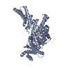
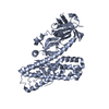
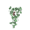
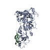
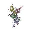
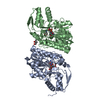
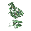
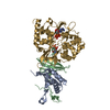
| Entry | Database: PDB / ID: 5wu6 | ||||||
|---|---|---|---|---|---|---|---|
| Title | Crystal structure of apo human Tut1, form IV | ||||||
 Components Components | Speckle targeted PIP5K1A-regulated poly(A) polymerase | ||||||
 Keywords Keywords | TRANSFERASE / Terminal nucleotidyl transferase | ||||||
| Function / homology |  Function and homology information Function and homology informationU6 snRNA 3'-end processing / RNA uridylyltransferase / co-transcriptional mRNA 3'-end processing, cleavage and polyadenylation pathway / regulation of RNA metabolic process / RNA uridylyltransferase activity / RNA 3'-end processing / snRNA processing / mRNA cleavage and polyadenylation specificity factor complex / polynucleotide adenylyltransferase / poly(A) RNA polymerase activity ...U6 snRNA 3'-end processing / RNA uridylyltransferase / co-transcriptional mRNA 3'-end processing, cleavage and polyadenylation pathway / regulation of RNA metabolic process / RNA uridylyltransferase activity / RNA 3'-end processing / snRNA processing / mRNA cleavage and polyadenylation specificity factor complex / polynucleotide adenylyltransferase / poly(A) RNA polymerase activity / mRNA 3'-end processing / enzyme-substrate adaptor activity / U6 snRNA binding / mRNA 3'-UTR binding / nuclear speck / nucleolus / enzyme binding / RNA binding / zinc ion binding / nucleoplasm / ATP binding / cytosol Similarity search - Function | ||||||
| Biological species |  Homo sapiens (human) Homo sapiens (human) | ||||||
| Method |  X-RAY DIFFRACTION / SYNCHROTRON / MOLECULAR REPLACEMENT / Resolution: 3.209 Å X-RAY DIFFRACTION / SYNCHROTRON / MOLECULAR REPLACEMENT / Resolution: 3.209 Å | ||||||
 Authors Authors | Yamashita, S. / Tomita, K. | ||||||
| Funding support |  Japan, 1items Japan, 1items
| ||||||
 Citation Citation | Journal: Nat Commun / Year: 2017 Title: Crystal structures of U6 snRNA-specific terminal uridylyltransferase Authors: Yamashita, S. / Takagi, Y. / Nagaike, T. / Tomita, K. | ||||||
| History |
|
- Structure visualization
| Structure viewer | Molecule: MolmilJmol/JSmol |
|---|
- Downloads & links
-Download
| PDBx/mmCIF format | 5wu6.cif.gz | 624.8 KB | Display | PDBx/mmCIF format |
|---|---|---|---|---|
| PDB format | pdb5wu6.ent.gz | 518.9 KB | Display | PDB format |
| PDBx/mmJSON format | 5wu6.json.gz | Tree view | PDBx/mmJSON format | |
| Others |  Other downloads Other downloads |
-Validation report
| Arichive directory | https://data.pdbj.org/pub/pdb/validation_reports/wu/5wu6ftp://data.pdbj.org/pub/pdb/validation_reports/wu/5wu6 | HTTPS FTP |
|---|
-Related structure data
| Related structure data | 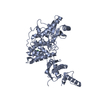 5wu1SC 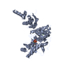 5wu2C 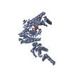 5wu3C 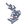 5wu4C 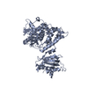 5wu5C C: citing same article ( S: Starting model for refinement |
|---|---|
| Similar structure data |

-Links
 PDBj
PDBj


- Assembly
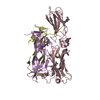
| Deposited unit | 
| ||||||||
|---|---|---|---|---|---|---|---|---|---|
| 1 | 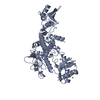
| ||||||||
| 2 | 
| ||||||||
| 3 | 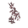
| ||||||||
| 4 | 
| ||||||||
| Unit cell |
|
-Components
| #1: Protein | Mass: 52828.816 Da / Num. of mol.: 4 / Fragment: UNP residues 54-599 / Mutation: C372A, C399A, C415A, C501A, C504A, C574A Source method: isolated from a genetically manipulated source Details: Sequence of 141-874(C-term) of human Tut1 was cloned to pET22b vector with NdeI and XhoI sites. Residues of 235-304 and 651-750 were deleted for crystallization. C372A, C399A, C415A, C501A, ...Details: Sequence of 141-874(C-term) of human Tut1 was cloned to pET22b vector with NdeI and XhoI sites. Residues of 235-304 and 651-750 were deleted for crystallization. C372A, C399A, C415A, C501A, C504S, and C574A mutations were introduced to improve the crystals. Source: (gene. exp.) Homo sapiens (human) / Gene: TUT1, RBM21 / Production host:  References: UniProt: Q9H6E5, polynucleotide adenylyltransferase, RNA uridylyltransferase #2: Chemical | ChemComp-MG /   Mass: 24.305 Da / Num. of mol.: 7 / Source method: obtained synthetically / Formula: Mg Mass: 24.305 Da / Num. of mol.: 7 / Source method: obtained synthetically / Formula: Mg |
|---|
-Experimental details
-Experiment
| Experiment | Method: X-RAY DIFFRACTION / Number of used crystals: 1 |
|---|
- Sample preparation
| Crystal | Density Matthews: 3.4 Å3/Da / Density % sol: 68.6 % |
|---|---|
| Crystal grow | Temperature: 277 K / Method: vapor diffusion, hanging drop / Details: Tris pH 7.0, PEG3350, MgCl2. |
-Data collection
| Diffraction | Mean temperature: 95 K | |||||||||||||||||||||||||||||||||||||||||||||||||||||||||||||||||||||||||||||||||||||||||||||||||||||||||||||||||||||||||||||||||||||||||||||||||||
|---|---|---|---|---|---|---|---|---|---|---|---|---|---|---|---|---|---|---|---|---|---|---|---|---|---|---|---|---|---|---|---|---|---|---|---|---|---|---|---|---|---|---|---|---|---|---|---|---|---|---|---|---|---|---|---|---|---|---|---|---|---|---|---|---|---|---|---|---|---|---|---|---|---|---|---|---|---|---|---|---|---|---|---|---|---|---|---|---|---|---|---|---|---|---|---|---|---|---|---|---|---|---|---|---|---|---|---|---|---|---|---|---|---|---|---|---|---|---|---|---|---|---|---|---|---|---|---|---|---|---|---|---|---|---|---|---|---|---|---|---|---|---|---|---|---|---|---|---|
| Diffraction source | Source: SYNCHROTRON / Site: Photon Factory / Beamline: BL-17A / Wavelength: 0.98 Å | |||||||||||||||||||||||||||||||||||||||||||||||||||||||||||||||||||||||||||||||||||||||||||||||||||||||||||||||||||||||||||||||||||||||||||||||||||
| Detector | Type: DECTRIS PILATUS3 S 6M / Detector: PIXEL / Date: Dec 7, 2015 | |||||||||||||||||||||||||||||||||||||||||||||||||||||||||||||||||||||||||||||||||||||||||||||||||||||||||||||||||||||||||||||||||||||||||||||||||||
| Radiation | Protocol: SINGLE WAVELENGTH / Monochromatic (M) / Laue (L): M / Scattering type: x-ray | |||||||||||||||||||||||||||||||||||||||||||||||||||||||||||||||||||||||||||||||||||||||||||||||||||||||||||||||||||||||||||||||||||||||||||||||||||
| Radiation wavelength | Wavelength: 0.98 Å / Relative weight: 1 | |||||||||||||||||||||||||||||||||||||||||||||||||||||||||||||||||||||||||||||||||||||||||||||||||||||||||||||||||||||||||||||||||||||||||||||||||||
| Reflection | Resolution: 3.2→50 Å / Num. obs: 48397 / % possible obs: 99.9 % / Observed criterion σ(I): -3 / Redundancy: 26.4 % / Biso Wilson estimate: 70.09 Å2 / CC1/2: 0.995 / Rmerge(I) obs: 0.375 / Net I/σ(I): 14.32 | |||||||||||||||||||||||||||||||||||||||||||||||||||||||||||||||||||||||||||||||||||||||||||||||||||||||||||||||||||||||||||||||||||||||||||||||||||
| Reflection shell |
|
- Processing
| Software |
| ||||||||||||||||||||||||||||||||||||||||||||||||||||||||||||||||||||||||||||||||||||||||||||||||||||||||||||||||||||||||||||||||||||||||||||||||||||||||||||||||||||||||||||||||||||||||||||||||||||||||
|---|---|---|---|---|---|---|---|---|---|---|---|---|---|---|---|---|---|---|---|---|---|---|---|---|---|---|---|---|---|---|---|---|---|---|---|---|---|---|---|---|---|---|---|---|---|---|---|---|---|---|---|---|---|---|---|---|---|---|---|---|---|---|---|---|---|---|---|---|---|---|---|---|---|---|---|---|---|---|---|---|---|---|---|---|---|---|---|---|---|---|---|---|---|---|---|---|---|---|---|---|---|---|---|---|---|---|---|---|---|---|---|---|---|---|---|---|---|---|---|---|---|---|---|---|---|---|---|---|---|---|---|---|---|---|---|---|---|---|---|---|---|---|---|---|---|---|---|---|---|---|---|---|---|---|---|---|---|---|---|---|---|---|---|---|---|---|---|---|---|---|---|---|---|---|---|---|---|---|---|---|---|---|---|---|---|---|---|---|---|---|---|---|---|---|---|---|---|---|---|---|---|
| Refinement | Method to determine structure: MOLECULAR REPLACEMENT Starting model: 5WU1 Resolution: 3.209→19.993 Å / SU ML: 0.35 / Cross valid method: FREE R-VALUE / σ(F): 1.36 / Phase error: 23.55
| ||||||||||||||||||||||||||||||||||||||||||||||||||||||||||||||||||||||||||||||||||||||||||||||||||||||||||||||||||||||||||||||||||||||||||||||||||||||||||||||||||||||||||||||||||||||||||||||||||||||||
| Solvent computation | Shrinkage radii: 0.9 Å / VDW probe radii: 1.11 Å | ||||||||||||||||||||||||||||||||||||||||||||||||||||||||||||||||||||||||||||||||||||||||||||||||||||||||||||||||||||||||||||||||||||||||||||||||||||||||||||||||||||||||||||||||||||||||||||||||||||||||
| Displacement parameters | Biso max: 223.09 Å2 / Biso mean: 73.73 Å2 / Biso min: 28.4 Å2 | ||||||||||||||||||||||||||||||||||||||||||||||||||||||||||||||||||||||||||||||||||||||||||||||||||||||||||||||||||||||||||||||||||||||||||||||||||||||||||||||||||||||||||||||||||||||||||||||||||||||||
| Refinement step | Cycle: final / Resolution: 3.209→19.993 Å
| ||||||||||||||||||||||||||||||||||||||||||||||||||||||||||||||||||||||||||||||||||||||||||||||||||||||||||||||||||||||||||||||||||||||||||||||||||||||||||||||||||||||||||||||||||||||||||||||||||||||||
| Refine LS restraints |
| ||||||||||||||||||||||||||||||||||||||||||||||||||||||||||||||||||||||||||||||||||||||||||||||||||||||||||||||||||||||||||||||||||||||||||||||||||||||||||||||||||||||||||||||||||||||||||||||||||||||||
| LS refinement shell | Refine-ID: X-RAY DIFFRACTION / Total num. of bins used: 17
| ||||||||||||||||||||||||||||||||||||||||||||||||||||||||||||||||||||||||||||||||||||||||||||||||||||||||||||||||||||||||||||||||||||||||||||||||||||||||||||||||||||||||||||||||||||||||||||||||||||||||
| Refinement TLS params. | Method: refined / Refine-ID: X-RAY DIFFRACTION
| ||||||||||||||||||||||||||||||||||||||||||||||||||||||||||||||||||||||||||||||||||||||||||||||||||||||||||||||||||||||||||||||||||||||||||||||||||||||||||||||||||||||||||||||||||||||||||||||||||||||||
| Refinement TLS group |
|
