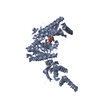
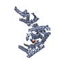
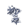
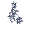
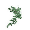
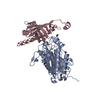
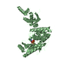
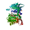
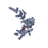
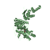
SpeckletargetedPIP5K1A-regulatedpoly(A) polymerase / Star-PAP / RNA-binding motif protein 21 / RNA-binding protein 21 / U6 snRNA-specific terminal ...Star-PAP / RNA-binding motif protein 21 / RNA-binding protein 21 / U6 snRNA-specific terminal uridylyltransferase 1 / U6-TUTase
Mass: 62317.711 Da / Num. of mol.: 2 / Fragment: UNP residues 141-874 / Mutation: C372A, C415A, C501A, C504S Source method: isolated from a genetically manipulated source Details: Sequence of 141-874(C-term) of human Tut1 was cloned to pET22b vector with NdeI and XhoI sites. Residues of 235-304 and 651-750 were deleted for crystallization. C372A, C415A, C501A, and ...Details: Sequence of 141-874(C-term) of human Tut1 was cloned to pET22b vector with NdeI and XhoI sites. Residues of 235-304 and 651-750 were deleted for crystallization. C372A, C415A, C501A, and C504S mutations were introduced to improve the crystals. Source: (gene. exp.) Homo sapiens (human) / Gene: TUT1, RBM21 / Production host: Escherichia coli (E. coli) References: UniProt: Q9H6E5, polynucleotide adenylyltransferase, RNA uridylyltransferase
In the structure databanks used in Yorodumi, some data are registered as the other names, "COVID-19 virus" and "2019-nCoV". Here are the details of the virus and the list of structure data.
Jan 31, 2019. EMDB accession codes are about to change! (news from PDBe EMDB page)
EMDB accession codes are about to change! (news from PDBe EMDB page)
The allocation of 4 digits for EMDB accession codes will soon come to an end. Whilst these codes will remain in use, new EMDB accession codes will include an additional digit and will expand incrementally as the available range of codes is exhausted. The current 4-digit format prefixed with “EMD-” (i.e. EMD-XXXX) will advance to a 5-digit format (i.e. EMD-XXXXX), and so on. It is currently estimated that the 4-digit codes will be depleted around Spring 2019, at which point the 5-digit format will come into force.
The EM Navigator/Yorodumi systems omit the EMD- prefix.
Related info.:Q: What is EMD? / ID/Accession-code notation in Yorodumi/EM Navigator
Yorodumi is a browser for structure data from EMDB, PDB, SASBDB, etc.
This page is also the successor to EM Navigator detail page, and also detail information page/front-end page for Omokage search.
The word "yorodu" (or yorozu) is an old Japanese word meaning "ten thousand". "mi" (miru) is to see.
Related info.:EMDB / PDB / SASBDB / Comparison of 3 databanks / Yorodumi Search / Aug 31, 2016. New EM Navigator & Yorodumi / Yorodumi Papers / Jmol/JSmol / Function and homology information / Changes in new EM Navigator and Yorodumi
 Movie
Movie Controller


 Open data
 Basic information
 Components
 Keywords
 Function and homology information
 Homo sapiens (human)
 X-RAY DIFFRACTION /
  Authors
 Japan, 1items
  Citation
 Structure visualization
 Downloads & links
 Other downloads
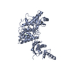


 PDBj
PDBj


 Assembly
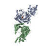


 Mass: 484.141 Da / Num. of mol.: 2 / Source method: obtained synthetically / Formula: C9H15N2O15P3 / Comment: UTP*YM
 Mass: 137.327 Da / Num. of mol.: 2 / Source method: obtained synthetically / Formula: Ba
 Mass: 35.453 Da / Num. of mol.: 2 / Source method: obtained synthetically / Formula: Cl
 Sample preparation
 Processing