 Movie
Movie Controller

[English] 日本語
 Yorodumi
- PDB-4c5h: Crystal structure of the minimal Pho-Sfmbt complex (P3121 spacegroup) -
+ Open data

- Basic information
| Entry | Database: PDB / ID: 4c5h | ||||||
|---|---|---|---|---|---|---|---|
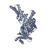
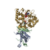
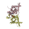
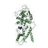
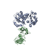
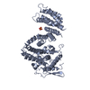
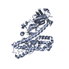
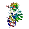
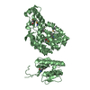
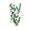
| Title | Crystal structure of the minimal Pho-Sfmbt complex (P3121 spacegroup) | ||||||
 Components Components |
| ||||||
 Keywords Keywords | TRANSCRIPTION | ||||||
| Function / homology |  Function and homology information Function and homology informationTranscriptional Regulation by E2F6 / histone H4K20me1 reader activity / polytene chromosome puff / DNA Damage Recognition in GG-NER / imaginal disc growth / SUMOylation of chromatin organization proteins / Estrogen-dependent gene expression / UCH proteinases / polytene chromosome / histone H4K20me2 reader activity ...Transcriptional Regulation by E2F6 / histone H4K20me1 reader activity / polytene chromosome puff / DNA Damage Recognition in GG-NER / imaginal disc growth / SUMOylation of chromatin organization proteins / Estrogen-dependent gene expression / UCH proteinases / polytene chromosome / histone H4K20me2 reader activity / PcG protein complex / histone H3K4me1 reader activity / Ino80 complex / dendrite morphogenesis / anterior/posterior axis specification / histone H3K9me2/3 reader activity / oogenesis / DNA topological change / transcription initiation-coupled chromatin remodeling / chromatin DNA binding / heterochromatin formation / DNA recombination / transcription regulator complex / sequence-specific DNA binding / DNA-binding transcription factor activity, RNA polymerase II-specific / RNA polymerase II cis-regulatory region sequence-specific DNA binding / chromatin remodeling / negative regulation of gene expression / negative regulation of DNA-templated transcription / DNA repair / chromatin binding / regulation of transcription by RNA polymerase II / regulation of DNA-templated transcription / chromatin / DNA binding / zinc ion binding / nucleus Similarity search - Function | ||||||
| Biological species |  | ||||||
| Method |  X-RAY DIFFRACTION / SYNCHROTRON / MOLECULAR REPLACEMENT / Resolution: 3.2 Å X-RAY DIFFRACTION / SYNCHROTRON / MOLECULAR REPLACEMENT / Resolution: 3.2 Å | ||||||
 Authors Authors | Alfieri, C. / Glatt, S. / Mueller, C.W. | ||||||
 Citation Citation | Journal: Genes Dev. / Year: 2013 Title: Structural Basis for Targeting the Chromatin Repressor Sfmbt to Polycomb Response Elements Authors: Alfieri, C. / Gambetta, M.C. / Matos, R. / Glatt, S. / Sehr, P. / Fraterman, S. / Wilm, M. / Mueller, J. / Mueller, C.W. | ||||||
| History |
|
- Structure visualization
| Structure viewer | Molecule: MolmilJmol/JSmol |
|---|
- Downloads & links
-Download
| PDBx/mmCIF format | 4c5h.cif.gz | 103.9 KB | Display | PDBx/mmCIF format |
|---|---|---|---|---|
| PDB format | pdb4c5h.ent.gz | 77.6 KB | Display | PDB format |
| PDBx/mmJSON format | 4c5h.json.gz | Tree view | PDBx/mmJSON format | |
| Others |  Other downloads Other downloads |
-Validation report
| Arichive directory | https://data.pdbj.org/pub/pdb/validation_reports/c5/4c5hftp://data.pdbj.org/pub/pdb/validation_reports/c5/4c5h | HTTPS FTP |
|---|
-Related structure data
| Related structure data |  4c5eC  4c5gC  4c5iC 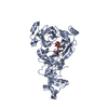 3h6zS C: citing same article ( S: Starting model for refinement |
|---|---|
| Similar structure data |
-Links
 PDBj
PDBj


- Assembly
| Deposited unit | 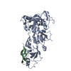
| ||||||||
|---|---|---|---|---|---|---|---|---|---|
| 1 |
| ||||||||
| Unit cell |
|
-Components
| #1: Protein | Mass: 51067.664 Da / Num. of mol.: 1 / Fragment: 4MBT, RESIDUES 531-980 Source method: isolated from a genetically manipulated source Source: (gene. exp.)  |
|---|---|
| #2: Protein | Mass: 15019.656 Da / Num. of mol.: 1 / Fragment: SPACER, RESIDUES 116-246 Source method: isolated from a genetically manipulated source Source: (gene. exp.) |
-Experimental details
-Experiment
| Experiment | Method: X-RAY DIFFRACTION |
|---|
- Sample preparation
| Crystal | Density Matthews: 4.88 Å3/Da / Density % sol: 80.3 % / Description: NONE |
|---|
-Data collection
| Diffraction | Mean temperature: 100 K |
|---|---|
| Diffraction source | Source: SYNCHROTRON / Site: ESRF  / Beamline: ID14-4 / Wavelength: 1.13747 / Beamline: ID14-4 / Wavelength: 1.13747 |
| Detector | Type: ADSC CCD / Detector: CCD / Date: Feb 17, 2011 |
| Radiation | Protocol: SINGLE WAVELENGTH / Monochromatic (M) / Laue (L): M / Scattering type: x-ray |
| Radiation wavelength | Wavelength: 1.13747 Å / Relative weight: 1 |
| Reflection | Resolution: 3.2→3.28 Å / Num. obs: 21760 / % possible obs: 99.9 % / Observed criterion σ(I): 1.5 / Redundancy: 7.7 % / Biso Wilson estimate: 85.62 Å2 / Rmerge(I) obs: 0.2 / Net I/σ(I): 10.3 |
| Reflection shell | Resolution: 3.2→3.28 Å / Redundancy: 7.6 % / Mean I/σ(I) obs: 1.3 / % possible all: 99.6 |
- Processing
| Software |
| |||||||||||||||||||||||||||||||||||||||||||||||||||||||||||||||
|---|---|---|---|---|---|---|---|---|---|---|---|---|---|---|---|---|---|---|---|---|---|---|---|---|---|---|---|---|---|---|---|---|---|---|---|---|---|---|---|---|---|---|---|---|---|---|---|---|---|---|---|---|---|---|---|---|---|---|---|---|---|---|---|---|
| Refinement | Method to determine structure: MOLECULAR REPLACEMENT Starting model: PDB ENTRY 3H6Z Resolution: 3.2→49.489 Å / SU ML: 0.49 / σ(F): 1.36 / Phase error: 26.22 / Stereochemistry target values: ML
| |||||||||||||||||||||||||||||||||||||||||||||||||||||||||||||||
| Solvent computation | Shrinkage radii: 0.9 Å / VDW probe radii: 1.11 Å / Solvent model: FLAT BULK SOLVENT MODEL | |||||||||||||||||||||||||||||||||||||||||||||||||||||||||||||||
| Displacement parameters | Biso mean: 46.2 Å2 | |||||||||||||||||||||||||||||||||||||||||||||||||||||||||||||||
| Refinement step | Cycle: LAST / Resolution: 3.2→49.489 Å
| |||||||||||||||||||||||||||||||||||||||||||||||||||||||||||||||
| Refine LS restraints |
| |||||||||||||||||||||||||||||||||||||||||||||||||||||||||||||||
| LS refinement shell |
|
