 Movie
Movie Controller

[English] 日本語
 Yorodumi
- PDB-6h5q: Cryo-EM structure of in vitro assembled Measles virus N into nucl... -
+ Open data

- Basic information
| Entry | Database: PDB / ID: 6h5q | ||||||||||||||||||
|---|---|---|---|---|---|---|---|---|---|---|---|---|---|---|---|---|---|---|---|
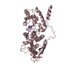
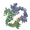
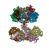
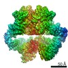
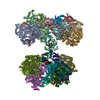
| Title | Cryo-EM structure of in vitro assembled Measles virus N into nucleocapsid-like particles (NCLPs) bound to polyA RNA hexamers. | ||||||||||||||||||
 Components Components |
| ||||||||||||||||||
 Keywords Keywords | VIRAL PROTEIN / Measles / Nucleocapsid / Helical / RNA | ||||||||||||||||||
| Function / homology |  Function and homology information Function and homology informationhelical viral capsid / viral nucleocapsid / host cell cytoplasm / ribonucleoprotein complex / host cell nucleus / structural molecule activity / RNA binding Similarity search - Function | ||||||||||||||||||
| Biological species |   Measles morbillivirus Measles morbillivirussynthetic construct (others) | ||||||||||||||||||
| Method | ELECTRON MICROSCOPY / helical reconstruction / cryo EM / Resolution: 3.3 Å | ||||||||||||||||||
 Authors Authors | Desfosses, A. / Milles, S. / Ringkjobing Jensen, M. / Guseva, S. / Colletier, J. / Maurin, D. / Schoehn, G. / Gutsche, I. / Ruigrok, R. / Blackledge, M. | ||||||||||||||||||
| Funding support |  France, 5items France, 5items
| ||||||||||||||||||
 Citation Citation | Journal: Proc Natl Acad Sci U S A / Year: 2019 Title: Assembly and cryo-EM structures of RNA-specific measles virus nucleocapsids provide mechanistic insight into paramyxoviral replication. Authors: Ambroise Desfosses / Sigrid Milles / Malene Ringkjøbing Jensen / Serafima Guseva / Jacques-Philippe Colletier / Damien Maurin / Guy Schoehn / Irina Gutsche / Rob W H Ruigrok / Martin Blackledge / Abstract: Assembly of paramyxoviral nucleocapsids on the RNA genome is an essential step in the viral cycle. The structural basis of this process has remained obscure due to the inability to control ...Assembly of paramyxoviral nucleocapsids on the RNA genome is an essential step in the viral cycle. The structural basis of this process has remained obscure due to the inability to control encapsidation. We used a recently developed approach to assemble measles virus nucleocapsid-like particles on specific sequences of RNA hexamers (poly-Adenine and viral genomic 5') in vitro, and determined their cryoelectron microscopy maps to 3.3-Å resolution. The structures unambiguously determine 5' and 3' binding sites and thereby the binding-register of viral genomic RNA within nucleocapsids. This observation reveals that the 3' end of the genome is largely exposed in fully assembled measles nucleocapsids. In particular, the final three nucleotides of the genome are rendered accessible to the RNA-dependent RNA polymerase complex, possibly enabling efficient RNA processing. The structures also reveal local and global conformational changes in the nucleoprotein upon assembly, in particular involving helix α6 and helix α13 that form edges of the RNA binding groove. Disorder is observed in the bound RNA, localized at one of the two backbone conformational switch sites. The high-resolution structure allowed us to identify putative nucleobase interaction sites in the RNA-binding groove, whose impact on assembly kinetics was measured using real-time NMR. Mutation of one of these sites, R195, whose sidechain stabilizes both backbone and base of a bound nucleic acid, is thereby shown to be essential for nucleocapsid-like particle assembly. | ||||||||||||||||||
| History |
|
- Structure visualization
| Movie |
Movie viewer |
|---|---|
| Structure viewer | Molecule: MolmilJmol/JSmol |
- Downloads & links
-Download
| PDBx/mmCIF format | 6h5q.cif.gz | 82.4 KB | Display | PDBx/mmCIF format |
|---|---|---|---|---|
| PDB format | pdb6h5q.ent.gz | 60.1 KB | Display | PDB format |
| PDBx/mmJSON format | 6h5q.json.gz | Tree view | PDBx/mmJSON format | |
| Others |  Other downloads Other downloads |
-Validation report
| Arichive directory | https://data.pdbj.org/pub/pdb/validation_reports/h5/6h5qftp://data.pdbj.org/pub/pdb/validation_reports/h5/6h5q | HTTPS FTP |
|---|
-Related structure data
| Related structure data | 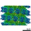 0141MC 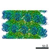 0142C 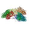 6h5sC M: map data used to model this data C: citing same article ( |
|---|---|
| Similar structure data |
-Links
 PDBj
PDBj

- Assembly
| Deposited unit | 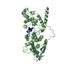
|
|---|---|
| 1 | x 15
|
-Components
| #1: Protein | Mass: 46425.863 Da / Num. of mol.: 1 Source method: isolated from a genetically manipulated source Source: (gene. exp.) Measles morbillivirus / Production host:  |
|---|---|
| #2: RNA chain | Mass: 1930.277 Da / Num. of mol.: 1 / Source method: obtained synthetically / Source: (synth.) synthetic construct (others) |
-Experimental details
-Experiment
| Experiment | Method: ELECTRON MICROSCOPY |
|---|---|
| EM experiment | Aggregation state: HELICAL ARRAY / 3D reconstruction method: helical reconstruction |
- Sample preparation
| Component |
| ||||||||||||||||||||||||
|---|---|---|---|---|---|---|---|---|---|---|---|---|---|---|---|---|---|---|---|---|---|---|---|---|---|
| Molecular weight | Units: KILODALTONS/NANOMETER / Experimental value: NO | ||||||||||||||||||||||||
| Source (natural) |
| ||||||||||||||||||||||||
| Source (recombinant) |
| ||||||||||||||||||||||||
| Buffer solution | pH: 6 / Details: 50 mM Na-phosphate pH 6, 150 mM NaCl, 2 mM DTT | ||||||||||||||||||||||||
| Specimen | Conc.: 1 mg/ml / Embedding applied: NO / Shadowing applied: NO / Staining applied: NO / Vitrification applied: YES | ||||||||||||||||||||||||
| Specimen support | Grid material: COPPER / Grid type: Quantifoil R2/1 | ||||||||||||||||||||||||
| Vitrification | Instrument: FEI VITROBOT MARK IV / Cryogen name: ETHANE / Humidity: 100 % / Chamber temperature: 293.15 K |
- Electron microscopy imaging
| Experimental equipment |  Model: Tecnai Polara / Image courtesy: FEI Company |
|---|---|
| Microscopy | Model: FEI POLARA 300 |
| Electron gun | Electron source:  FIELD EMISSION GUN / Accelerating voltage: 300 kV / Illumination mode: FLOOD BEAM FIELD EMISSION GUN / Accelerating voltage: 300 kV / Illumination mode: FLOOD BEAM |
| Electron lens | Mode: BRIGHT FIELD / Nominal magnification: 23000 X / Nominal defocus max: 3 nm / Nominal defocus min: 0.8 nm / Cs: 2 mm / Alignment procedure: COMA FREE |
| Image recording | Average exposure time: 4 sec. / Electron dose: 30 e/Å2 / Detector mode: SUPER-RESOLUTION / Film or detector model: GATAN K2 SUMMIT (4k x 4k) / Num. of grids imaged: 1 / Num. of real images: 186 |
| Image scans | Sampling size: 5 µm / Width: 4000 / Height: 4000 / Movie frames/image: 40 / Used frames/image: 2-20 |
- Processing
| EM software |
| ||||||||||||||||||||||||||||
|---|---|---|---|---|---|---|---|---|---|---|---|---|---|---|---|---|---|---|---|---|---|---|---|---|---|---|---|---|---|
| CTF correction | Type: PHASE FLIPPING AND AMPLITUDE CORRECTION | ||||||||||||||||||||||||||||
| Helical symmerty | Angular rotation/subunit: -29.17105583 ° / Axial rise/subunit: 3.920265781 Å / Axial symmetry: C1 | ||||||||||||||||||||||||||||
| Particle selection | Num. of particles selected: 111582 | ||||||||||||||||||||||||||||
| 3D reconstruction | Resolution: 3.3 Å / Resolution method: FSC 0.143 CUT-OFF / Num. of particles: 111582 / Algorithm: BACK PROJECTION / Symmetry type: HELICAL | ||||||||||||||||||||||||||||
| Refinement | Highest resolution: 3.3 Å |
