 Movie
Movie Controller


+ Open data

- Basic information
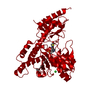
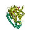
| Entry | Database: PDB / ID: 5wt4 | ||||||||||||
|---|---|---|---|---|---|---|---|---|---|---|---|---|---|
| Title | L-Cysteine-PLP intermediate of NifS from Helicobacter pylori | ||||||||||||
 Components Components | Cysteine desulfurase IscS | ||||||||||||
 Keywords Keywords | TRANSFERASE / Iron-sulfur cluster biogenesis / cysteine desulfurase | ||||||||||||
| Function / homology |  Function and homology information Function and homology informationcysteine desulfurase / cysteine desulfurase activity / : / [2Fe-2S] cluster assembly / 2 iron, 2 sulfur cluster binding / pyridoxal phosphate binding / metal ion binding / cytoplasm Similarity search - Function | ||||||||||||
| Biological species |   Helicobacter pylori (bacteria) Helicobacter pylori (bacteria) | ||||||||||||
| Method |  X-RAY DIFFRACTION / SYNCHROTRON / MOLECULAR REPLACEMENT / Resolution: 2.92 Å X-RAY DIFFRACTION / SYNCHROTRON / MOLECULAR REPLACEMENT / Resolution: 2.92 Å | ||||||||||||
 Authors Authors | Fujishiro, T. / Nakamura, R. / Takahashi, T. | ||||||||||||
| Funding support |  Japan, 3items Japan, 3items
| ||||||||||||
 Citation Citation | Journal: Febs J. / Year: 2019 Title: Snapshots of PLP-substrate and PLP-product external aldimines as intermediates in two types of cysteine desulfurase enzymes. Authors: Nakamura, R. / Hikita, M. / Ogawa, S. / Takahashi, Y. / Fujishiro, T. | ||||||||||||
| History |
|
- Structure visualization
| Structure viewer | Molecule: MolmilJmol/JSmol |
|---|
- Downloads & links
-Download
| PDBx/mmCIF format | 5wt4.cif.gz | 159.1 KB | Display | PDBx/mmCIF format |
|---|---|---|---|---|
| PDB format | pdb5wt4.ent.gz | 124.6 KB | Display | PDB format |
| PDBx/mmJSON format | 5wt4.json.gz | Tree view | PDBx/mmJSON format | |
| Others |  Other downloads Other downloads |
-Validation report
| Arichive directory | https://data.pdbj.org/pub/pdb/validation_reports/wt/5wt4ftp://data.pdbj.org/pub/pdb/validation_reports/wt/5wt4 | HTTPS FTP |
|---|
-Related structure data
| Related structure data |  5wt2C  5zs9C  5zskC  5zsoC  5zspC  5zstC  6kfzC  6kg0C  6kg1C  1ecxS C: citing same article ( S: Starting model for refinement |
|---|---|
| Similar structure data |


-Links
 PDBj
PDBj
- Assembly
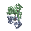
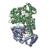
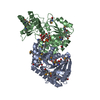
| Deposited unit | 
| ||||||||
|---|---|---|---|---|---|---|---|---|---|
| 1 | 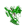
| ||||||||
| Unit cell |
|
-Components
| #1: Protein | Mass: 44125.230 Da / Num. of mol.: 1 / Mutation: L2V, K138R Source method: isolated from a genetically manipulated source Source: (gene. exp.) Helicobacter pylori (strain ATCC 700392 / 26695) (bacteria)Strain: ATCC 700392 / 26695 / Gene: iscS, HP_0220 / Production host: |
|---|---|
| #2: Chemical | ChemComp-IPA /   Mass: 60.095 Da / Num. of mol.: 1 / Source method: obtained synthetically / Formula: C3H8O Mass: 60.095 Da / Num. of mol.: 1 / Source method: obtained synthetically / Formula: C3H8O |
| #3: Chemical | ChemComp-C6P /   Mass: 352.301 Da / Num. of mol.: 1 / Source method: obtained synthetically / Formula: C11H17N2O7PS Mass: 352.301 Da / Num. of mol.: 1 / Source method: obtained synthetically / Formula: C11H17N2O7PS |
| #4: Water | ChemComp-HOH /  Mass: 18.015 Da / Num. of mol.: 9 / Source method: isolated from a natural source / Formula: H2O Mass: 18.015 Da / Num. of mol.: 9 / Source method: isolated from a natural source / Formula: H2O |
-Experimental details
-Experiment
| Experiment | Method: X-RAY DIFFRACTION / Number of used crystals: 1 |
|---|
- Sample preparation
| Crystal | Density Matthews: 4.03 Å3/Da / Density % sol: 69.47 % |
|---|---|
| Crystal grow | Temperature: 293 K / Method: vapor diffusion, sitting drop / pH: 7.5 / Details: HEPES/NaOH, PEG4000, glycerol, isopropanol |
-Data collection
| Diffraction | Mean temperature: 100 K |
|---|---|
| Diffraction source | Source: SYNCHROTRON / Site: SPring-8 / Beamline: BL38B1 / Wavelength: 1 Å |
| Detector | Type: ADSC QUANTUM 315r / Detector: CCD / Date: Jun 3, 2016 |
| Radiation | Monochromator: Si (111) double crystal monochromator / Protocol: SINGLE WAVELENGTH / Monochromatic (M) / Laue (L): M / Scattering type: x-ray |
| Radiation wavelength | Wavelength: 1 Å / Relative weight: 1 |
| Reflection | Resolution: 2.92→50 Å / Num. obs: 16036 / % possible obs: 97.8 % / Redundancy: 13.3 % / Biso Wilson estimate: 66.6 Å2 / CC1/2: 0.997 / Rmerge(I) obs: 0.1082 / Net I/σ(I): 26.52 |
| Reflection shell | Resolution: 2.92→3.024 Å / Redundancy: 14.5 % / Rmerge(I) obs: 0.9236 / Mean I/σ(I) obs: 3.59 / Num. unique all: 1579 / CC1/2: 0.908 / % possible all: 99.8 |
- Processing
| Software |
| ||||||||||||||||||||||||||||||||||||||||||||||||||||||||||||||||||||||||||||||||||||||||||||||||||||||||||||||||||||||||||||||||||||||||||||||||||||||
|---|---|---|---|---|---|---|---|---|---|---|---|---|---|---|---|---|---|---|---|---|---|---|---|---|---|---|---|---|---|---|---|---|---|---|---|---|---|---|---|---|---|---|---|---|---|---|---|---|---|---|---|---|---|---|---|---|---|---|---|---|---|---|---|---|---|---|---|---|---|---|---|---|---|---|---|---|---|---|---|---|---|---|---|---|---|---|---|---|---|---|---|---|---|---|---|---|---|---|---|---|---|---|---|---|---|---|---|---|---|---|---|---|---|---|---|---|---|---|---|---|---|---|---|---|---|---|---|---|---|---|---|---|---|---|---|---|---|---|---|---|---|---|---|---|---|---|---|---|---|---|---|
| Refinement | Method to determine structure: MOLECULAR REPLACEMENT Starting model: 1ECX Resolution: 2.92→48.123 Å / SU ML: 0.34 / Cross valid method: FREE R-VALUE / σ(F): 1.36 / Phase error: 32.07
| ||||||||||||||||||||||||||||||||||||||||||||||||||||||||||||||||||||||||||||||||||||||||||||||||||||||||||||||||||||||||||||||||||||||||||||||||||||||
| Solvent computation | Shrinkage radii: 0.9 Å / VDW probe radii: 1.11 Å | ||||||||||||||||||||||||||||||||||||||||||||||||||||||||||||||||||||||||||||||||||||||||||||||||||||||||||||||||||||||||||||||||||||||||||||||||||||||
| Refinement step | Cycle: LAST / Resolution: 2.92→48.123 Å
| ||||||||||||||||||||||||||||||||||||||||||||||||||||||||||||||||||||||||||||||||||||||||||||||||||||||||||||||||||||||||||||||||||||||||||||||||||||||
| Refine LS restraints |
| ||||||||||||||||||||||||||||||||||||||||||||||||||||||||||||||||||||||||||||||||||||||||||||||||||||||||||||||||||||||||||||||||||||||||||||||||||||||
| LS refinement shell | Refine-ID: X-RAY DIFFRACTION
| ||||||||||||||||||||||||||||||||||||||||||||||||||||||||||||||||||||||||||||||||||||||||||||||||||||||||||||||||||||||||||||||||||||||||||||||||||||||
| Refinement TLS params. | Method: refined / Refine-ID: X-RAY DIFFRACTION
| ||||||||||||||||||||||||||||||||||||||||||||||||||||||||||||||||||||||||||||||||||||||||||||||||||||||||||||||||||||||||||||||||||||||||||||||||||||||
| Refinement TLS group |
|
