[English] 日本語
Yorodumi
- PDB-3ly1: Crystal structure of Putative histidinol-phosphate aminotransfera... -

+
Open data


ID or keywords:

Loading...

-
Basic information

Entry
Database: PDB / ID: 3ly1
TitleCrystal structure of Putative histidinol-phosphate aminotransferase (YP_050345.1) from Erwinia carotovora atroseptica SCRI1043 at 1.80 A resolution
ComponentsPutative histidinol-phosphate aminotransferase
KeywordsTRANSFERASE / Putative histidinol-phosphate aminotransferase / Structural Genomics / Joint Center for Structural Genomics / JCSG / Protein Structure Initiative / PSI-2 / Aminotransferase
Function / homology
Function and homology information


transaminase activity / biosynthetic process / pyridoxal phosphate binding
Similarity search - Function
Aminotransferase, class I/classII / Aminotransferase class I and II / Aspartate Aminotransferase, domain 1 / Twin arginine translocation (Tat) signal profile. / Twin-arginine translocation pathway, signal sequence / Aspartate Aminotransferase, domain 1 / Aspartate Aminotransferase; domain 2 / Type I PLP-dependent aspartate aminotransferase-like (Major domain) / Pyridoxal phosphate-dependent transferase, small domain / Pyridoxal phosphate-dependent transferase, major domain ...Aminotransferase, class I/classII / Aminotransferase class I and II / Aspartate Aminotransferase, domain 1 / Twin arginine translocation (Tat) signal profile. / Twin-arginine translocation pathway, signal sequence / Aspartate Aminotransferase, domain 1 / Aspartate Aminotransferase; domain 2 / Type I PLP-dependent aspartate aminotransferase-like (Major domain) / Pyridoxal phosphate-dependent transferase, small domain / Pyridoxal phosphate-dependent transferase, major domain / Pyridoxal phosphate-dependent transferase / Alpha-Beta Complex / 3-Layer(aba) Sandwich / Alpha Beta
Similarity search - Domain/homology
CITRIC ACID / PYRIDOXAL-5'-PHOSPHATE / Putative aminotransferase
Similarity search - Component
Biological speciesErwinia carotovora atroseptica (bacteria)
MethodX-RAY DIFFRACTION / SYNCHROTRON / MAD / Resolution: 1.8 Å
AuthorsJoint Center for Structural Genomics (JCSG)
CitationJournal: To be published
Title: Crystal structure of Putative histidinol-phosphate aminotransferase (YP_050345.1) from Erwinia carotovora atroseptica SCRI1043 at 1.80 A resolution
Authors: Joint Center for Structural Genomics (JCSG)
History
DepositionFeb 25, 2010Deposition site: RCSB / Processing site: RCSB
Revision 1.0Mar 31, 2010Provider: repository / Type: Initial release
Revision 1.1Jul 13, 2011Group: Advisory / Version format compliance
Revision 1.2Nov 8, 2017Group: Refinement description / Category: software
Revision 1.3Jul 17, 2019Group: Data collection / Derived calculations / Refinement description
Category: software / struct_conn
Item: _software.classification / _software.contact_author ..._software.classification / _software.contact_author / _software.contact_author_email / _software.location / _software.name / _software.type / _software.version / _struct_conn.pdbx_leaving_atom_flag
Revision 1.4Feb 1, 2023Group: Database references / Derived calculations / Category: database_2 / struct_ref_seq_dif / struct_site
Item: _database_2.pdbx_DOI / _database_2.pdbx_database_accession ..._database_2.pdbx_DOI / _database_2.pdbx_database_accession / _struct_ref_seq_dif.details / _struct_site.pdbx_auth_asym_id / _struct_site.pdbx_auth_comp_id / _struct_site.pdbx_auth_seq_id

-
Structure visualization

Structure viewerMolecule:
MolmilJmol/JSmol

Downloads & links

-
Assembly

Deposited unit
A: Putative histidinol-phosphate aminotransferase
B: Putative histidinol-phosphate aminotransferase
C: Putative histidinol-phosphate aminotransferase
D: Putative histidinol-phosphate aminotransferase
hetero molecules


Theoretical massNumber of molelcules
Total (without water)160,12435
Polymers156,9394
Non-polymers3,18531
Water32,2831792
1
A: Putative histidinol-phosphate aminotransferase
B: Putative histidinol-phosphate aminotransferase
hetero molecules


Theoretical massNumber of molelcules
Total (without water)79,90715
Polymers78,4702
Non-polymers1,43713
Water362
TypeNameSymmetry operationNumber
identity operation1_555x,y,z1
Buried area8350 Å2
ΔGint25 kcal/mol
Surface area26000 Å2
MethodPISA
2
C: Putative histidinol-phosphate aminotransferase
D: Putative histidinol-phosphate aminotransferase
hetero molecules


Theoretical massNumber of molelcules
Total (without water)80,21720
Polymers78,4702
Non-polymers1,74718
Water362
TypeNameSymmetry operationNumber
identity operation1_555x,y,z1
Buried area8280 Å2
ΔGint20 kcal/mol
Surface area26810 Å2
MethodPISA
Unit cell
Length a, b, c (Å)58.625, 150.477, 100.092
Angle α, β, γ (deg.)90.000, 94.380, 90.000
Int Tables number4
Space group name H-MP1211
DetailsCRYSTAL PACKING ANALYSIS SUGGESTS THE ASSIGNMENT OF A DIMER AS THE SIGNIFICANT OLIGOMERIZATION STATE.

-
Components

#1: Protein
Putative histidinol-phosphate aminotransferase


Mass: 39234.832 Da / Num. of mol.: 4 / Fragment: sequence database residues 42-494
Source method: isolated from a genetically manipulated source
Source: (gene. exp.) Erwinia carotovora atroseptica (bacteria)
Gene: ECA2250 / Plasmid: SpeedET / Production host: Escherichia coli (E. coli) / Strain (production host): HK100 / References: UniProt: Q6D4Z0
#2: Chemical
ChemComp-PLP / PYRIDOXAL-5'-PHOSPHATE / VITAMIN B6 Phosphate / Pyridoxal phosphate


Mass: 247.142 Da / Num. of mol.: 4 / Source method: obtained synthetically / Formula: C8H10NO6P
#3: Chemical
ChemComp-CIT / CITRIC ACID / Citric acid


Mass: 192.124 Da / Num. of mol.: 4 / Source method: obtained synthetically / Formula: C6H8O7
#4: Chemical...
ChemComp-EDO / 1,2-ETHANEDIOL / ETHYLENE GLYCOL / Ethylene glycol


Mass: 62.068 Da / Num. of mol.: 23 / Source method: obtained synthetically / Formula: C2H6O2
#5: Water ChemComp-HOH / water / Water


Mass: 18.015 Da / Num. of mol.: 1792 / Source method: isolated from a natural source / Formula: H2O
Sequence details
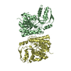
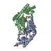
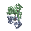
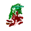
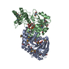
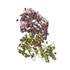
THIS CONSTRUCT (RESIDUES 42-394 OF THE FULL LENGTH TARGET) WAS EXPRESSED WITH A PURIFICATION TAG ...THIS CONSTRUCT (RESIDUES 42-394 OF THE FULL LENGTH TARGET) WAS EXPRESSED WITH A PURIFICATION TAG MGSDKIHHHHHHENLYFQG. THE TAG WAS REMOVED WITH TEV PROTEASE LEAVING ONLY A GLYCINE (0) FOLLOWED BY THE TARGET SEQUENCE.

-
Experimental details

-
Experiment

ExperimentMethod: X-RAY DIFFRACTION / Number of used crystals: 1

-
Sample preparation

CrystalDensity Matthews: 2.8 Å3/Da / Density % sol: 56.15 %
Crystal growTemperature: 277 K / Method: vapor diffusion, sitting drop / pH: 5.6
Details: 20.0000% iso-Propanol, 20.0000% PEG-4000, 0.1M Citrate pH 5.6, NANODROP, VAPOR DIFFUSION, SITTING DROP, temperature 277K

-
Data collection

DiffractionMean temperature: 100 K
Diffraction sourceSource: SYNCHROTRON / Site: SSRL / Beamline: BL9-2 / Wavelength: 0.96109,0.97946,0.97936
DetectorType: MARMOSAIC 325 mm CCD / Detector: CCD / Date: Nov 7, 2009 / Details: Flat collimating mirror, toroid focusing mirror
RadiationMonochromator: Double crystal monochromator / Protocol: MAD / Monochromatic (M) / Laue (L): M / Scattering type: x-ray
Radiation wavelength
IDWavelength (Å)Relative weight
10.961091
20.979461
30.979361
ReflectionResolution: 1.8→29.564 Å / Num. obs: 158547 / % possible obs: 96.5 % / Observed criterion σ(I): -3 / Biso Wilson estimate: 23.011 Å2 / Rmerge(I) obs: 0.059 / Net I/σ(I): 8.36
Reflection shell
Resolution (Å)Rmerge(I) obsMean I/σ(I) obsNum. measured obsNum. unique obsDiffraction-ID% possible all
1.79-1.850.6751.44422624110179.8
1.85-1.930.5221.86490334301198.7
1.93-2.020.3472.66164932378198.7
2.02-2.120.2453.55680529711198.6
2.12-2.250.1794.65995831250198.5
2.25-2.430.1216.46333432937198.7
2.43-2.670.0918.16017731209198.6
2.67-3.060.05811.46161331886198.2
3.06-3.850.03218.16048131235197.4
3.85-29.5640.02224.76108031348197

-
Phasing

PhasingMethod: MAD

-
Processing

Software
NameVersionClassificationNB
REFMAC5.5.0102refinement
PHENIXrefinement
SHELXphasing
XSCALEdata scaling
PDB_EXTRACT3.006data extraction
XDSdata reduction
SHELXDphasing
MolProbity3beta29model building
RefinementMethod to determine structure: MAD / Resolution: 1.8→29.564 Å / Cor.coef. Fo:Fc: 0.972 / Cor.coef. Fo:Fc free: 0.961 / Occupancy max: 1 / Occupancy min: 0.15 / SU B: 6.336 / SU ML: 0.086 / TLS residual ADP flag: LIKELY RESIDUAL / Cross valid method: THROUGHOUT / σ(F): 0 / ESU R: 0.118 / ESU R Free: 0.111
Stereochemistry target values: MAXIMUM LIKELIHOOD WITH PHASES
Details: 1. HYDROGENS HAVE BEEN ADDED IN THE RIDING POSITIONS. 2. ATOM RECORDS CONTAIN RESIDUAL B FACTORS ONLY. 3. A MET-INHIBITION PROTOCOL WAS USED FOR SELENOMETHIONINE INCORPORATION DURING PROTEIN ...Details: 1. HYDROGENS HAVE BEEN ADDED IN THE RIDING POSITIONS. 2. ATOM RECORDS CONTAIN RESIDUAL B FACTORS ONLY. 3. A MET-INHIBITION PROTOCOL WAS USED FOR SELENOMETHIONINE INCORPORATION DURING PROTEIN EXPRESSION. THE OCCUPANCY OF THE SE ATOMS IN THE MSE RESIDUES WAS REDUCED TO 0.75 FOR THE REDUCED SCATTERING POWER DUE TO PARTIAL S-MET INCORPORATION. 4. CITRATE (CIT) AND ETHYLENE GLYCOL (EDO) MOLECULES FROM THE CRYSTALLIZATION/CRYOPROTECTION SOLUTION ARE MODELED. 5. PYRIDOXAL-5'-PHOSPHATE (PLP) IS MODELED NEAR LYS-253 ON THE BASIS OF ELECTRON DENSITY AND HOMOLOGY TO OTHER PLP DEPENDANT ENZYMES.
RfactorNum. reflection% reflectionSelection details
Rfree0.206 7952 5 %RANDOM
Rwork0.176 ---
obs0.178 158407 99.32 %-
Solvent computationIon probe radii: 0.8 Å / Shrinkage radii: 0.8 Å / VDW probe radii: 1.4 Å / Solvent model: BABINET MODEL WITH MASK
Displacement parametersBiso max: 77.81 Å2 / Biso mean: 17.717 Å2 / Biso min: 2 Å2
Baniso -1Baniso -2Baniso -3
1-0.66 Å20 Å2-1.05 Å2
2---3.33 Å20 Å2
3---2.51 Å2
Refinement stepCycle: LAST / Resolution: 1.8→29.564 Å
ProteinNucleic acidLigandSolventTotal
Num. atoms10683 0 208 1792 12683
Refine LS restraints
Refine-IDTypeDev idealDev ideal targetNumber
X-RAY DIFFRACTIONr_bond_refined_d0.0170.02211535
X-RAY DIFFRACTIONr_bond_other_d0.0010.027800
X-RAY DIFFRACTIONr_angle_refined_deg1.5331.97215721
X-RAY DIFFRACTIONr_angle_other_deg0.946319140
X-RAY DIFFRACTIONr_dihedral_angle_1_deg6.05351506
X-RAY DIFFRACTIONr_dihedral_angle_2_deg31.05524.295475
X-RAY DIFFRACTIONr_dihedral_angle_3_deg12.806151927
X-RAY DIFFRACTIONr_dihedral_angle_4_deg17.8131560
X-RAY DIFFRACTIONr_chiral_restr0.0890.21743
X-RAY DIFFRACTIONr_gen_planes_refined0.0070.02112847
X-RAY DIFFRACTIONr_gen_planes_other0.0010.022238
X-RAY DIFFRACTIONr_nbd_refined
X-RAY DIFFRACTIONr_nbd_other
X-RAY DIFFRACTIONr_nbtor_refined
X-RAY DIFFRACTIONr_nbtor_other
X-RAY DIFFRACTIONr_xyhbond_nbd_refined
X-RAY DIFFRACTIONr_xyhbond_nbd_other
X-RAY DIFFRACTIONr_metal_ion_refined
X-RAY DIFFRACTIONr_metal_ion_other
X-RAY DIFFRACTIONr_symmetry_vdw_refined
X-RAY DIFFRACTIONr_symmetry_vdw_other
X-RAY DIFFRACTIONr_symmetry_hbond_refined
X-RAY DIFFRACTIONr_symmetry_hbond_other
X-RAY DIFFRACTIONr_symmetry_metal_ion_refined
X-RAY DIFFRACTIONr_symmetry_metal_ion_other
X-RAY DIFFRACTIONr_mcbond_it0.7721.57162
X-RAY DIFFRACTIONr_mcbond_other0.2541.52846
X-RAY DIFFRACTIONr_mcangle_it1.26821159
X-RAY DIFFRACTIONr_scbond_it2.22834373
X-RAY DIFFRACTIONr_scangle_it3.4634.54073
X-RAY DIFFRACTIONr_rigid_bond_restr
X-RAY DIFFRACTIONr_sphericity_free
X-RAY DIFFRACTIONr_sphericity_bonded
LS refinement shellResolution: 1.8→1.847 Å / Total num. of bins used: 20
RfactorNum. reflection% reflection
Rfree0.322 576 -
Rwork0.293 10846 -
all-11422 -
obs--97.27 %
Refinement TLS params.

Method: refined / Refine-ID: X-RAY DIFFRACTION

IDL112)L122)L132)L222)L232)L332)S11 (Å °)S12 (Å °)S13 (Å °)S21 (Å °)S22 (Å °)S23 (Å °)S31 (Å °)S32 (Å °)S33 (Å °)T112)T122)T132)T222)T232)T332)Origin x (Å)Origin y (Å)Origin z (Å)
10.2562-0.0714-0.06050.99990.08360.45340.0069-0.02050.02150.0817-0.02340.1266-0.06270.01640.01660.0978-0.00030.01840.1487-0.00260.04272.4547.691-49.89
20.3343-0.02010.02340.9411-0.12910.42710.0138-0.0037-0.04980.0344-0.0217-0.01130.08180.04340.00790.12490.01750.00860.15240.0180.020381.90113.119-49.919
30.1942-0.0786-0.12371.3179-0.12840.26480.04990.03220.0116-0.1071-0.0433-0.1239-0.0565-0.0076-0.00660.1120.01540.06660.19810.0150.045348.77267.31-0.087
40.28760.15720.07551.36520.28920.38720.01640.0232-0.0097-0.0525-0.05750.05630.0691-0.04960.04110.1037-0.0040.04890.1945-0.01140.031239.0532.715-0.577
Refinement TLS group
IDRefine-IDRefine TLS-IDAuth asym-IDAuth seq-ID
1X-RAY DIFFRACTION1A49 - 394
2X-RAY DIFFRACTION2B49 - 394
3X-RAY DIFFRACTION3C48 - 394
4X-RAY DIFFRACTION4D0 - 394

+
About Yorodumi

-
News

-
Feb 9, 2022. New format data for meta-information of EMDB entries

New format data for meta-information of EMDB entries

  • Version 3 of the EMDB header file is now the official format.
  • The previous official version 1.9 will be removed from the archive.

Related info.:EMDB header

External links:wwPDB to switch to version 3 of the EMDB data model

-
Aug 12, 2020. Covid-19 info

Covid-19 info

URL: https://pdbj.org/emnavi/covid19.php

New page: Covid-19 featured information page in EM Navigator.

Related info.:Covid-19 info / Mar 5, 2020. Novel coronavirus structure data

+
Mar 5, 2020. Novel coronavirus structure data

Novel coronavirus structure data

Related info.:Yorodumi Speices / Aug 12, 2020. Covid-19 info

External links:COVID-19 featured content - PDBj / Molecule of the Month (242):Coronavirus Proteases

+
Jan 31, 2019. EMDB accession codes are about to change! (news from PDBe EMDB page)

EMDB accession codes are about to change! (news from PDBe EMDB page)

  • The allocation of 4 digits for EMDB accession codes will soon come to an end. Whilst these codes will remain in use, new EMDB accession codes will include an additional digit and will expand incrementally as the available range of codes is exhausted. The current 4-digit format prefixed with “EMD-” (i.e. EMD-XXXX) will advance to a 5-digit format (i.e. EMD-XXXXX), and so on. It is currently estimated that the 4-digit codes will be depleted around Spring 2019, at which point the 5-digit format will come into force.
  • The EM Navigator/Yorodumi systems omit the EMD- prefix.

Related info.:Q: What is EMD? / ID/Accession-code notation in Yorodumi/EM Navigator

External links:EMDB Accession Codes are Changing Soon! / Contact to PDBj

+
Jul 12, 2017. Major update of PDB

Major update of PDB

  • wwPDB released updated PDB data conforming to the new PDBx/mmCIF dictionary.
  • This is a major update changing the version number from 4 to 5, and with Remediation, in which all the entries are updated.
  • In this update, many items about electron microscopy experimental information are reorganized (e.g. em_software).
  • Now, EM Navigator and Yorodumi are based on the updated data.

External links:wwPDB Remediation / Enriched Model Files Conforming to OneDep Data Standards Now Available in the PDB FTP Archive

-
Yorodumi

Thousand views of thousand structures

  • Yorodumi is a browser for structure data from EMDB, PDB, SASBDB, etc.
  • This page is also the successor to EM Navigator detail page, and also detail information page/front-end page for Omokage search.
  • The word "yorodu" (or yorozu) is an old Japanese word meaning "ten thousand". "mi" (miru) is to see.

Related info.:EMDB / PDB / SASBDB / Comparison of 3 databanks / Yorodumi Search / Aug 31, 2016. New EM Navigator & Yorodumi / Yorodumi Papers / Jmol/JSmol / Function and homology information / Changes in new EM Navigator and Yorodumi

Read more