 Movie
Movie Controller


+ Open data

- Basic information
| Entry | Database: PDB / ID: 5w9q | ||||||
|---|---|---|---|---|---|---|---|
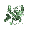
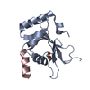
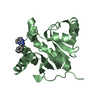
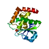
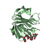
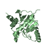
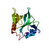
| Title | Zinc finger region of MBD1 in complex with CpG DNA | ||||||
 Components Components |
| ||||||
 Keywords Keywords | DNA BINDING PROTEIN/DNA / zinc finger / dna-binding / CXXC3 / Structural Genomics / Structural Genomics Consortium / SGC / DNA BINDING PROTEIN-DNA complex | ||||||
| Function / homology |  Function and homology information Function and homology informationdouble-stranded methylated DNA binding / unmethylated CpG binding / methyl-CpG binding / DNA methylation-dependent constitutive heterochromatin formation / SUMOylation of transcription cofactors / nuclear matrix / chromosome / transcription by RNA polymerase II / nuclear speck / negative regulation of DNA-templated transcription ...double-stranded methylated DNA binding / unmethylated CpG binding / methyl-CpG binding / DNA methylation-dependent constitutive heterochromatin formation / SUMOylation of transcription cofactors / nuclear matrix / chromosome / transcription by RNA polymerase II / nuclear speck / negative regulation of DNA-templated transcription / negative regulation of transcription by RNA polymerase II / DNA binding / zinc ion binding / nucleoplasm / nucleus Similarity search - Function | ||||||
| Biological species |  Homo sapiens (human) Homo sapiens (human)synthetic construct (others) | ||||||
| Method |  X-RAY DIFFRACTION / SYNCHROTRON / MOLECULAR REPLACEMENT / Resolution: 1.8 Å X-RAY DIFFRACTION / SYNCHROTRON / MOLECULAR REPLACEMENT / Resolution: 1.8 Å | ||||||
 Authors Authors | Liu, K. / Xu, C. / Tempel, W. / Walker, J.R. / Arrowsmith, C.H. / Bountra, C. / Edwards, A.M. / Min, J. / Structural Genomics Consortium (SGC) | ||||||
 Citation Citation | Journal: Structure / Year: 2018 Title: DNA Sequence Recognition of Human CXXC Domains and Their Structural Determinants. Authors: Xu, C. / Liu, K. / Lei, M. / Yang, A. / Li, Y. / Hughes, T.R. / Min, J. | ||||||
| History |
|
- Structure visualization
| Structure viewer | Molecule: MolmilJmol/JSmol |
|---|
- Downloads & links
-Download
| PDBx/mmCIF format | 5w9q.cif.gz | 113.6 KB | Display | PDBx/mmCIF format |
|---|---|---|---|---|
| PDB format | pdb5w9q.ent.gz | 84.2 KB | Display | PDB format |
| PDBx/mmJSON format | 5w9q.json.gz | Tree view | PDBx/mmJSON format | |
| Others |  Other downloads Other downloads |
-Validation report
| Arichive directory | https://data.pdbj.org/pub/pdb/validation_reports/w9/5w9qftp://data.pdbj.org/pub/pdb/validation_reports/w9/5w9q | HTTPS FTP |
|---|
-Related structure data
| Related structure data |  4nw3C  4o64C  4pziC 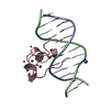 4z3cC  5vc9C  5w9sC  6asbC  6asdC  3qmdS 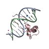 4hp3S S: Starting model for refinement C: citing same article ( |
|---|---|
| Similar structure data |


-Links
 PDBj
PDBj


- Assembly
| Deposited unit | 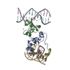
| ||||||||
|---|---|---|---|---|---|---|---|---|---|
| 1 | 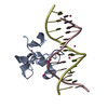
| ||||||||
| 2 | 
| ||||||||
| Unit cell |
|
-Components
| #1: Protein | Mass: 7003.344 Da / Num. of mol.: 2 / Fragment: Zinc finger region (UNP residues 330-388) Source method: isolated from a genetically manipulated source Source: (gene. exp.) Homo sapiens (human) / Gene: MBD1, CXXC3, PCM1 / Plasmid: pET28-MHL / Production host:  #2: DNA chain | Mass: 3663.392 Da / Num. of mol.: 4 / Source method: obtained synthetically / Source: (synth.) synthetic construct (others) #3: Chemical | ChemComp-ZN /   Mass: 65.409 Da / Num. of mol.: 4 / Source method: obtained synthetically / Formula: Zn Mass: 65.409 Da / Num. of mol.: 4 / Source method: obtained synthetically / Formula: Zn#4: Chemical | ChemComp-UNX /   Num. of mol.: 16 / Source method: obtained synthetically Num. of mol.: 16 / Source method: obtained synthetically#5: Water | ChemComp-HOH / |  Mass: 18.015 Da / Num. of mol.: 46 / Source method: isolated from a natural source / Formula: H2O Mass: 18.015 Da / Num. of mol.: 46 / Source method: isolated from a natural source / Formula: H2O |
|---|
-Experimental details
-Experiment
| Experiment | Method: X-RAY DIFFRACTION / Number of used crystals: 1 |
|---|
- Sample preparation
| Crystal | Density Matthews: 2.2 Å3/Da / Density % sol: 43.4 % |
|---|---|
| Crystal grow | Temperature: 291 K / Method: vapor diffusion / pH: 7.5 / Details: 25% PEG-400, 0.2 M magnesium chloride, 0.1 M hepes |
-Data collection
| Diffraction | Mean temperature: 100 K | ||||||||||||||||||||||||||||||
|---|---|---|---|---|---|---|---|---|---|---|---|---|---|---|---|---|---|---|---|---|---|---|---|---|---|---|---|---|---|---|---|
| Diffraction source | Source: SYNCHROTRON / Site: CLSI  / Beamline: 08ID-1 / Wavelength: 0.97959 Å / Beamline: 08ID-1 / Wavelength: 0.97959 Å | ||||||||||||||||||||||||||||||
| Detector | Type: RAYONIX MX-300 / Detector: CCD / Date: Jun 1, 2013 | ||||||||||||||||||||||||||||||
| Radiation | Protocol: SINGLE WAVELENGTH / Monochromatic (M) / Laue (L): M / Scattering type: x-ray | ||||||||||||||||||||||||||||||
| Radiation wavelength | Wavelength: 0.97959 Å / Relative weight: 1 | ||||||||||||||||||||||||||||||
| Reflection | Resolution: 1.8→33.69 Å / Num. obs: 22639 / % possible obs: 99.8 % / Redundancy: 3.7 % / CC1/2: 0.998 / Rmerge(I) obs: 0.046 / Rpim(I) all: 0.028 / Rrim(I) all: 0.054 / Net I/σ(I): 13.3 / Num. measured all: 83217 / Scaling rejects: 0 | ||||||||||||||||||||||||||||||
| Reflection shell | Diffraction-ID: 1
|
- Processing
| Software |
| |||||||||||||||||||||||||||||||||||||||||||||||||||||||||||||||||||||||||||||||||||||||||||||||||||||||||||||||||||||||||||||||||||||||||||||||||||||||||||||||||||||||||||||||
|---|---|---|---|---|---|---|---|---|---|---|---|---|---|---|---|---|---|---|---|---|---|---|---|---|---|---|---|---|---|---|---|---|---|---|---|---|---|---|---|---|---|---|---|---|---|---|---|---|---|---|---|---|---|---|---|---|---|---|---|---|---|---|---|---|---|---|---|---|---|---|---|---|---|---|---|---|---|---|---|---|---|---|---|---|---|---|---|---|---|---|---|---|---|---|---|---|---|---|---|---|---|---|---|---|---|---|---|---|---|---|---|---|---|---|---|---|---|---|---|---|---|---|---|---|---|---|---|---|---|---|---|---|---|---|---|---|---|---|---|---|---|---|---|---|---|---|---|---|---|---|---|---|---|---|---|---|---|---|---|---|---|---|---|---|---|---|---|---|---|---|---|---|---|---|---|---|
| Refinement | Method to determine structure: MOLECULAR REPLACEMENT Starting model: PDB entries 4HP3, 3QMD Resolution: 1.8→33.69 Å / Cor.coef. Fo:Fc: 0.961 / Cor.coef. Fo:Fc free: 0.951 / SU B: 11.831 / SU ML: 0.158 / Cross valid method: THROUGHOUT / σ(F): 0 / ESU R: 0.155 / ESU R Free: 0.139 Details: arp/warp was used in map improvement mode. coot was used for interactive model building. Model geometry was assessed on the molprobity server.
| |||||||||||||||||||||||||||||||||||||||||||||||||||||||||||||||||||||||||||||||||||||||||||||||||||||||||||||||||||||||||||||||||||||||||||||||||||||||||||||||||||||||||||||||
| Solvent computation | Ion probe radii: 0.8 Å / Shrinkage radii: 0.8 Å / VDW probe radii: 1.2 Å | |||||||||||||||||||||||||||||||||||||||||||||||||||||||||||||||||||||||||||||||||||||||||||||||||||||||||||||||||||||||||||||||||||||||||||||||||||||||||||||||||||||||||||||||
| Displacement parameters | Biso max: 109.41 Å2 / Biso mean: 48.642 Å2 / Biso min: 25.03 Å2
| |||||||||||||||||||||||||||||||||||||||||||||||||||||||||||||||||||||||||||||||||||||||||||||||||||||||||||||||||||||||||||||||||||||||||||||||||||||||||||||||||||||||||||||||
| Refinement step | Cycle: final / Resolution: 1.8→33.69 Å
| |||||||||||||||||||||||||||||||||||||||||||||||||||||||||||||||||||||||||||||||||||||||||||||||||||||||||||||||||||||||||||||||||||||||||||||||||||||||||||||||||||||||||||||||
| Refine LS restraints |
| |||||||||||||||||||||||||||||||||||||||||||||||||||||||||||||||||||||||||||||||||||||||||||||||||||||||||||||||||||||||||||||||||||||||||||||||||||||||||||||||||||||||||||||||
| LS refinement shell | Resolution: 1.8→1.847 Å / Total num. of bins used: 20
| |||||||||||||||||||||||||||||||||||||||||||||||||||||||||||||||||||||||||||||||||||||||||||||||||||||||||||||||||||||||||||||||||||||||||||||||||||||||||||||||||||||||||||||||
| Refinement TLS params. | Method: refined / Refine-ID: X-RAY DIFFRACTION
| |||||||||||||||||||||||||||||||||||||||||||||||||||||||||||||||||||||||||||||||||||||||||||||||||||||||||||||||||||||||||||||||||||||||||||||||||||||||||||||||||||||||||||||||
| Refinement TLS group |
|
