 Movie
Movie Controller


+ Open data

- Basic information
| Entry | Database: PDB / ID: 4pzi | ||||||
|---|---|---|---|---|---|---|---|
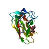
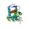
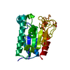
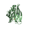
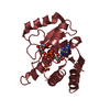
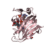
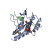
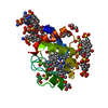
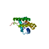
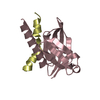
| Title | Zinc finger region of MLL2 in complex with CpG DNA | ||||||
 Components Components |
| ||||||
 Keywords Keywords | TRANSCRIPTION/DNA / zinc finger / dna-binding / Structural Genomics / Structural Genomics Consortium / SGC / TRANSCRIPTION-DNA complex | ||||||
| Function / homology |  Function and homology information Function and homology information[histone H3]-lysine4 N-methyltransferase / histone H3K4 monomethyltransferase activity / unmethylated CpG binding / MLL1/2 complex / histone H3K4 methyltransferase activity / Formation of WDR5-containing histone-modifying complexes / histone methyltransferase complex / RUNX1 regulates genes involved in megakaryocyte differentiation and platelet function / PKMTs methylate histone lysines / methylation ...[histone H3]-lysine4 N-methyltransferase / histone H3K4 monomethyltransferase activity / unmethylated CpG binding / MLL1/2 complex / histone H3K4 methyltransferase activity / Formation of WDR5-containing histone-modifying complexes / histone methyltransferase complex / RUNX1 regulates genes involved in megakaryocyte differentiation and platelet function / PKMTs methylate histone lysines / methylation / positive regulation of DNA-templated transcription / zinc ion binding / nucleoplasm / nucleus Similarity search - Function | ||||||
| Biological species |  Homo sapiens (human) Homo sapiens (human) | ||||||
| Method |  X-RAY DIFFRACTION / SYNCHROTRON / MOLECULAR REPLACEMENT / molecular replacement / Resolution: 2.15 Å X-RAY DIFFRACTION / SYNCHROTRON / MOLECULAR REPLACEMENT / molecular replacement / Resolution: 2.15 Å | ||||||
 Authors Authors | Chao, X. / Tempel, W. / Liu, K. / Dong, A. / Bountra, C. / Weigelt, J. / Arrowsmith, C.H. / Edwards, A.M. / Min, J. / Structural Genomics Consortium (SGC) | ||||||
 Citation Citation | Journal: Structure / Year: 2018 Title: DNA Sequence Recognition of Human CXXC Domains and Their Structural Determinants. Authors: Xu, C. / Liu, K. / Lei, M. / Yang, A. / Li, Y. / Hughes, T.R. / Min, J. | ||||||
| History |
|
- Structure visualization
| Structure viewer | Molecule: MolmilJmol/JSmol |
|---|
- Downloads & links
-Download
| PDBx/mmCIF format | 4pzi.cif.gz | 65.2 KB | Display | PDBx/mmCIF format |
|---|---|---|---|---|
| PDB format | pdb4pzi.ent.gz | 45.6 KB | Display | PDB format |
| PDBx/mmJSON format | 4pzi.json.gz | Tree view | PDBx/mmJSON format | |
| Others |  Other downloads Other downloads |
-Validation report
| Arichive directory | https://data.pdbj.org/pub/pdb/validation_reports/pz/4pziftp://data.pdbj.org/pub/pdb/validation_reports/pz/4pzi | HTTPS FTP |
|---|
-Related structure data
| Related structure data |  4nw3C  4o64C 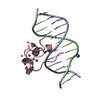 4z3cC  5vc9C  5w9qC  5w9sC  6asbC  6asdC C: citing same article ( |
|---|---|
| Similar structure data |
-Links
 PDBj
PDBj


- Assembly
| Deposited unit | 
| ||||||||
|---|---|---|---|---|---|---|---|---|---|
| 1 |
| ||||||||
| Unit cell |
| ||||||||
| Details | biological unit has not been determined |
-Components
| #1: Protein | Mass: 7563.124 Da / Num. of mol.: 1 Source method: isolated from a genetically manipulated source Source: (gene. exp.) Homo sapiens (human) / Gene: KMT2B, HRX2, KIAA0304, MLL2, MLL4, TRX2, WBP7 / Plasmid: pET28-MHL / Production host:  References: UniProt: Q9UMN6, histone-lysine N-methyltransferase | ||||||
|---|---|---|---|---|---|---|---|
| #2: DNA chain | Mass: 3664.380 Da / Num. of mol.: 2 / Source method: obtained synthetically / Details: synthetic dna #3: Chemical |   Mass: 65.409 Da / Num. of mol.: 2 / Source method: obtained synthetically / Formula: Zn Mass: 65.409 Da / Num. of mol.: 2 / Source method: obtained synthetically / Formula: Zn#4: Chemical | ChemComp-UNX / |   Num. of mol.: 1 / Source method: obtained synthetically Num. of mol.: 1 / Source method: obtained synthetically#5: Water | ChemComp-HOH / |  Mass: 18.015 Da / Num. of mol.: 8 / Source method: isolated from a natural source / Formula: H2O Mass: 18.015 Da / Num. of mol.: 8 / Source method: isolated from a natural source / Formula: H2O |
-Experimental details
-Experiment
| Experiment | Method: X-RAY DIFFRACTION / Number of used crystals: 1 |
|---|
- Sample preparation
| Crystal | Density Matthews: 2.8 Å3/Da / Density % sol: 55.7 % |
|---|---|
| Crystal grow | Temperature: 291 K / pH: 8.5 Details: 25% PEG-3350, 0.2 M ammonium sulfate, 0.1 M TRIS, pH 8.5, temperature 291K |
-Data collection
| Diffraction | Mean temperature: 100 K | |||||||||||||||||||||
|---|---|---|---|---|---|---|---|---|---|---|---|---|---|---|---|---|---|---|---|---|---|---|
| Diffraction source | Source: SYNCHROTRON / Site: APS  / Beamline: 19-ID / Wavelength: 0.97918 Å / Beamline: 19-ID / Wavelength: 0.97918 Å | |||||||||||||||||||||
| Detector | Type: ADSC QUANTUM 315 / Detector: CCD / Date: Aug 16, 2013 | |||||||||||||||||||||
| Radiation | Protocol: SINGLE WAVELENGTH / Monochromatic (M) / Laue (L): M / Scattering type: x-ray | |||||||||||||||||||||
| Radiation wavelength | Wavelength: 0.97918 Å / Relative weight: 1 | |||||||||||||||||||||
| Reflection | Resolution: 2.15→35.36 Å / Num. obs: 8659 / % possible obs: 99.8 % / Redundancy: 3.7 % / Rmerge(I) obs: 0.066 / Net I/σ(I): 14.3 | |||||||||||||||||||||
| Reflection shell | Diffraction-ID: 1
|
-Phasing
| Phasing | Method: molecular replacement |
|---|
- Processing
| Software |
| ||||||||||||||||||||||||||||||||||||||||||||||||||||||||||||||||||||||||||||||||||||||||||||||||||||
|---|---|---|---|---|---|---|---|---|---|---|---|---|---|---|---|---|---|---|---|---|---|---|---|---|---|---|---|---|---|---|---|---|---|---|---|---|---|---|---|---|---|---|---|---|---|---|---|---|---|---|---|---|---|---|---|---|---|---|---|---|---|---|---|---|---|---|---|---|---|---|---|---|---|---|---|---|---|---|---|---|---|---|---|---|---|---|---|---|---|---|---|---|---|---|---|---|---|---|---|---|---|
| Refinement | Method to determine structure: MOLECULAR REPLACEMENT Starting model: structure of isomorphous crystal was solved by molecular replacement using currently unpublished models of same protein and DNA, respectively. Resolution: 2.15→34.91 Å / Cor.coef. Fo:Fc: 0.947 / Cor.coef. Fo:Fc free: 0.939 / WRfactor Rfree: 0.2545 / WRfactor Rwork: 0.206 / Occupancy max: 1 / Occupancy min: 0.5 / FOM work R set: 0.7123 / SU B: 15.511 / SU ML: 0.204 / SU R Cruickshank DPI: 0.2259 / SU Rfree: 0.2013 / Cross valid method: THROUGHOUT / ESU R: 0.226 / ESU R Free: 0.201 / Stereochemistry target values: MAXIMUM LIKELIHOOD Details: coot was used for interactive model building. Model geometry was assessed with molprobity. I note the poor side chain fit of His-957 to electron density. I attempted modeling an alternate ...Details: coot was used for interactive model building. Model geometry was assessed with molprobity. I note the poor side chain fit of His-957 to electron density. I attempted modeling an alternate conformation in which N-terminal extension of main chain and this side chain are switched, but abandoned the attempt after failing to fit a good rotamer to the density.
| ||||||||||||||||||||||||||||||||||||||||||||||||||||||||||||||||||||||||||||||||||||||||||||||||||||
| Solvent computation | Ion probe radii: 0.8 Å / Shrinkage radii: 0.8 Å / VDW probe radii: 1.2 Å / Solvent model: MASK | ||||||||||||||||||||||||||||||||||||||||||||||||||||||||||||||||||||||||||||||||||||||||||||||||||||
| Displacement parameters | Biso max: 125.63 Å2 / Biso mean: 61.8311 Å2 / Biso min: 30.32 Å2
| ||||||||||||||||||||||||||||||||||||||||||||||||||||||||||||||||||||||||||||||||||||||||||||||||||||
| Refinement step | Cycle: LAST / Resolution: 2.15→34.91 Å
| ||||||||||||||||||||||||||||||||||||||||||||||||||||||||||||||||||||||||||||||||||||||||||||||||||||
| Refine LS restraints |
| ||||||||||||||||||||||||||||||||||||||||||||||||||||||||||||||||||||||||||||||||||||||||||||||||||||
| LS refinement shell | Resolution: 2.15→2.206 Å / Total num. of bins used: 20
| ||||||||||||||||||||||||||||||||||||||||||||||||||||||||||||||||||||||||||||||||||||||||||||||||||||
| Refinement TLS params. | Method: refined / Refine-ID: X-RAY DIFFRACTION
| ||||||||||||||||||||||||||||||||||||||||||||||||||||||||||||||||||||||||||||||||||||||||||||||||||||
| Refinement TLS group |
|
