- PDB-1e29: PSII associated cytochrome C549 from Synechocystis sp. -
+
Open data
ID or keywords:
Loading...
-
Basic information
Entry
Database: PDB / ID: 1.0E+29
Title
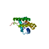
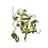
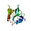
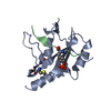
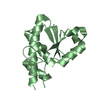
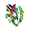
PSII associated cytochrome C549 from Synechocystis sp.
Components
CYTOCHROME C549
Keywords
ELECTRON TRANSPORT / PSII ASSOCIATED CYTOCHROME / CYTOCHROME / LOW POTENTIAL / BIS_HISTIDINYL / PSII MODULATOR
Function / homology
Function and homology information
plasma membrane-derived thylakoid photosystem II / plasma membrane-derived thylakoid membrane / photosynthesis, light reaction / respiratory electron transport chain / electron transfer activity / iron ion binding / heme binding Similarity search - Function
Mass: 18.015 Da / Num. of mol.: 223 / Source method: isolated from a natural source / Formula: H2O
Has protein modification
Y
-
Experimental details
-
Experiment
Experiment
Method: X-RAY DIFFRACTION / Number of used crystals: 1
-
Sample preparation
Crystal
Density Matthews: 1.99 Å3/Da / Density % sol: 48.7 % Description: LOCALIZATION OF FE ATOM BY PATTERSON ANALYSIS OF SUPER- SHARPENED NATIVE OR ANOMALOUS DIFFERENCE MAPS, FOLLOWED BY FE- ANCHORED ROTATION SEARCH OF CYS-GLY-GLY-CYS-HIS+HEME FRAGMENT AND ...Description: LOCALIZATION OF FE ATOM BY PATTERSON ANALYSIS OF SUPER- SHARPENED NATIVE OR ANOMALOUS DIFFERENCE MAPS, FOLLOWED BY FE- ANCHORED ROTATION SEARCH OF CYS-GLY-GLY-CYS-HIS+HEME FRAGMENT AND EXPANSION OF THIS SEED FRAGMENT TO FINAL SOLUTION
Crystal grow
Method: vapor diffusion, sitting drop / pH: 8.5 Details: SITTING DROP, VAPOUR DIFFUSION, 16% PEG 8000, O.2 M CALCIUM ACETATE, 0.1 M TRIS/HCL, PH = 8.5
In the structure databanks used in Yorodumi, some data are registered as the other names, "COVID-19 virus" and "2019-nCoV". Here are the details of the virus and the list of structure data.
Jan 31, 2019. EMDB accession codes are about to change! (news from PDBe EMDB page)
EMDB accession codes are about to change! (news from PDBe EMDB page)
The allocation of 4 digits for EMDB accession codes will soon come to an end. Whilst these codes will remain in use, new EMDB accession codes will include an additional digit and will expand incrementally as the available range of codes is exhausted. The current 4-digit format prefixed with “EMD-” (i.e. EMD-XXXX) will advance to a 5-digit format (i.e. EMD-XXXXX), and so on. It is currently estimated that the 4-digit codes will be depleted around Spring 2019, at which point the 5-digit format will come into force.
The EM Navigator/Yorodumi systems omit the EMD- prefix.
Related info.:Q: What is EMD? / ID/Accession-code notation in Yorodumi/EM Navigator
Yorodumi is a browser for structure data from EMDB, PDB, SASBDB, etc.
This page is also the successor to EM Navigator detail page, and also detail information page/front-end page for Omokage search.
The word "yorodu" (or yorozu) is an old Japanese word meaning "ten thousand". "mi" (miru) is to see.
Related info.:EMDB / PDB / SASBDB / Comparison of 3 databanks / Yorodumi Search / Aug 31, 2016. New EM Navigator & Yorodumi / Yorodumi Papers / Jmol/JSmol / Function and homology information / Changes in new EM Navigator and Yorodumi
 Movie
Movie Controller


 Open data
 Basic information
 Components
 Keywords
 Function and homology information
 X-RAY DIFFRACTION /
  Authors
 Citation
 Structure visualization
 Downloads & links
 Other downloads


 PDBj
PDBj


 Assembly
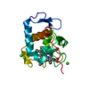


 Mass: 618.503 Da / Num. of mol.: 1 / Source method: obtained synthetically / Formula: C34H34FeN4O4
 Mass: 40.078 Da / Num. of mol.: 3 / Source method: obtained synthetically / Formula: Ca
 Mass: 18.015 Da / Num. of mol.: 223 / Source method: isolated from a natural source / Formula: H2O
 Sample preparation
 / Beamline: BW7B / Wavelength: 0.8374
  Processing