 Movie
Movie Controller


+ Open data

- Basic information
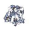
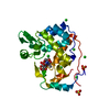
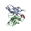
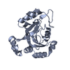
| Entry | Database: PDB / ID: 5uwa | ||||||||||||
|---|---|---|---|---|---|---|---|---|---|---|---|---|---|
| Title | Structure of E. coli phospholipid binding protein MlaC | ||||||||||||
 Components Components | Probable phospholipid-binding protein MlaC | ||||||||||||
 Keywords Keywords | TRANSPORT PROTEIN / phospholipid-binding protein / bacterial lipid transport | ||||||||||||
| Function / homology | Tgt2/MlaC superfamily / Toluene tolerance Ttg2/phospholipid-binding protein MlaC / MlaC protein / intermembrane phospholipid transfer / phospholipid transport / outer membrane-bounded periplasmic space / Chem-8ND / Intermembrane phospholipid transport system binding protein MlaC Function and homology information Function and homology information | ||||||||||||
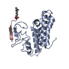
| Biological species |  | ||||||||||||
| Method |  X-RAY DIFFRACTION / SYNCHROTRON / MOLECULAR REPLACEMENT / Resolution: 1.501 Å X-RAY DIFFRACTION / SYNCHROTRON / MOLECULAR REPLACEMENT / Resolution: 1.501 Å | ||||||||||||
 Authors Authors | Bhabha, G. / Ekiert, D.C. | ||||||||||||
| Funding support |  United States, 3items United States, 3items
| ||||||||||||
 Citation Citation | Journal: Cell / Year: 2017 Title: Architectures of Lipid Transport Systems for the Bacterial Outer Membrane. Authors: Damian C Ekiert / Gira Bhabha / Georgia L Isom / Garrett Greenan / Sergey Ovchinnikov / Ian R Henderson / Jeffery S Cox / Ronald D Vale /  Abstract: How phospholipids are trafficked between the bacterial inner and outer membranes through the hydrophilic space of the periplasm is not known. We report that members of the mammalian cell entry (MCE) ...How phospholipids are trafficked between the bacterial inner and outer membranes through the hydrophilic space of the periplasm is not known. We report that members of the mammalian cell entry (MCE) protein family form hexameric assemblies with a central channel capable of mediating lipid transport. The E. coli MCE protein, MlaD, forms a ring associated with an ABC transporter complex in the inner membrane. A soluble lipid-binding protein, MlaC, ferries lipids between MlaD and an outer membrane protein complex. In contrast, EM structures of two other E. coli MCE proteins show that YebT forms an elongated tube consisting of seven stacked MCE rings, and PqiB adopts a syringe-like architecture. Both YebT and PqiB create channels of sufficient length to span the periplasmic space. This work reveals diverse architectures of highly conserved protein-based channels implicated in the transport of lipids between the membranes of bacteria and some eukaryotic organelles. | ||||||||||||
| History |
|
- Structure visualization
| Structure viewer | Molecule: MolmilJmol/JSmol |
|---|
- Downloads & links
-Download
| PDBx/mmCIF format | 5uwa.cif.gz | 186.5 KB | Display | PDBx/mmCIF format |
|---|---|---|---|---|
| PDB format | pdb5uwa.ent.gz | 148.4 KB | Display | PDB format |
| PDBx/mmJSON format | 5uwa.json.gz | Tree view | PDBx/mmJSON format | |
| Others |  Other downloads Other downloads |
-Validation report
| Arichive directory | https://data.pdbj.org/pub/pdb/validation_reports/uw/5uwaftp://data.pdbj.org/pub/pdb/validation_reports/uw/5uwa | HTTPS FTP |
|---|
-Related structure data
| Related structure data |  8608C  8610C  8611C  8612C 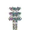 5uvnC 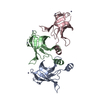 5uw2C 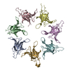 5uw8C 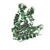 5uwbC  2qguS C: citing same article ( S: Starting model for refinement |
|---|---|
| Similar structure data |


-Links
 PDBj
PDBj- Assembly
| Deposited unit | 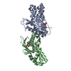
| ||||||||
|---|---|---|---|---|---|---|---|---|---|
| 1 | 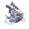
| ||||||||
| 2 | 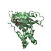
| ||||||||
| Unit cell |
|
-Components
| #1: Protein | Mass: 23512.613 Da / Num. of mol.: 2 Source method: isolated from a genetically manipulated source Source: (gene. exp.) Strain: K12 / Gene: mlaC, yrbC, b3192, JW3159 / Production host: #2: Chemical |   Mass: 611.979 Da / Num. of mol.: 2 / Source method: obtained synthetically / Formula: C37H73NO5 Mass: 611.979 Da / Num. of mol.: 2 / Source method: obtained synthetically / Formula: C37H73NO5#3: Water | ChemComp-HOH / |  Mass: 18.015 Da / Num. of mol.: 289 / Source method: isolated from a natural source / Formula: H2O Mass: 18.015 Da / Num. of mol.: 289 / Source method: isolated from a natural source / Formula: H2O |
|---|
-Experimental details
-Experiment
| Experiment | Method: X-RAY DIFFRACTION / Number of used crystals: 1 |
|---|
- Sample preparation
| Crystal | Density Matthews: 2.76 Å3/Da / Density % sol: 55.44 % |
|---|---|
| Crystal grow | Temperature: 295 K / Method: vapor diffusion, sitting drop Details: 0.1 M citric acid pH 3.5 and 1.6 M ammonium sulfate |
-Data collection
| Diffraction | Mean temperature: 100 K |
|---|---|
| Diffraction source | Source: SYNCHROTRON / Site: ALS / Beamline: 8.3.1 / Wavelength: 1.116 Å |
| Detector | Type: ADSC QUANTUM 315 / Detector: CCD / Date: May 21, 2015 |
| Radiation | Protocol: SINGLE WAVELENGTH / Monochromatic (M) / Laue (L): M / Scattering type: x-ray |
| Radiation wavelength | Wavelength: 1.116 Å / Relative weight: 1 |
| Reflection | Resolution: 1.5→50 Å / Num. obs: 83295 / % possible obs: 98.5 % / Redundancy: 7.2 % / CC1/2: 0.57 / Net I/σ(I): 22.2 |
- Processing
| Software |
| |||||||||||||||||||||||||||||||||||||||||||||||||||||||||||||||||||||||||||||||||||||||||||||||||||||||||
|---|---|---|---|---|---|---|---|---|---|---|---|---|---|---|---|---|---|---|---|---|---|---|---|---|---|---|---|---|---|---|---|---|---|---|---|---|---|---|---|---|---|---|---|---|---|---|---|---|---|---|---|---|---|---|---|---|---|---|---|---|---|---|---|---|---|---|---|---|---|---|---|---|---|---|---|---|---|---|---|---|---|---|---|---|---|---|---|---|---|---|---|---|---|---|---|---|---|---|---|---|---|---|---|---|---|---|
| Refinement | Method to determine structure: MOLECULAR REPLACEMENT Starting model: based on homology model with 2QGU as a template Resolution: 1.501→43.718 Å / SU ML: 0.16 / Cross valid method: FREE R-VALUE / σ(F): 0 / Phase error: 17.02
| |||||||||||||||||||||||||||||||||||||||||||||||||||||||||||||||||||||||||||||||||||||||||||||||||||||||||
| Solvent computation | Shrinkage radii: 0.9 Å / VDW probe radii: 1.2 Å | |||||||||||||||||||||||||||||||||||||||||||||||||||||||||||||||||||||||||||||||||||||||||||||||||||||||||
| Refinement step | Cycle: LAST / Resolution: 1.501→43.718 Å
| |||||||||||||||||||||||||||||||||||||||||||||||||||||||||||||||||||||||||||||||||||||||||||||||||||||||||
| Refine LS restraints |
| |||||||||||||||||||||||||||||||||||||||||||||||||||||||||||||||||||||||||||||||||||||||||||||||||||||||||
| LS refinement shell |
|
