 Movie
Movie Controller


+ Open data

- Basic information
| Entry | Database: EMDB / ID: EMD-8610 | ||||||||||||
|---|---|---|---|---|---|---|---|---|---|---|---|---|---|
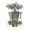
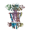
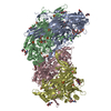
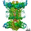
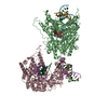
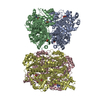
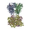
| Title | Structure of E. coli MCE protein PqiB, periplasmic domain | ||||||||||||
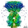
 Map data Map data | E. coli MCE protein PqiB, periplasmic domain | ||||||||||||
 Sample Sample |
| ||||||||||||
| Function / homology | : / Mce/MlaD / MlaD protein / intermembrane lipid transfer / membrane organization / outer membrane-bounded periplasmic space / identical protein binding / plasma membrane / Intermembrane transport protein PqiB Function and homology information Function and homology information | ||||||||||||
| Biological species |  | ||||||||||||
| Method | single particle reconstruction / cryo EM / Resolution: 10.0 Å | ||||||||||||
 Authors Authors | Bhabha G / Ekiert DC | ||||||||||||
| Funding support |  United States, 3 items United States, 3 items
| ||||||||||||
 Citation Citation | Journal: Cell / Year: 2017 Title: Architectures of Lipid Transport Systems for the Bacterial Outer Membrane. Authors: Damian C Ekiert / Gira Bhabha / Georgia L Isom / Garrett Greenan / Sergey Ovchinnikov / Ian R Henderson / Jeffery S Cox / Ronald D Vale /  Abstract: How phospholipids are trafficked between the bacterial inner and outer membranes through the hydrophilic space of the periplasm is not known. We report that members of the mammalian cell entry (MCE) ...How phospholipids are trafficked between the bacterial inner and outer membranes through the hydrophilic space of the periplasm is not known. We report that members of the mammalian cell entry (MCE) protein family form hexameric assemblies with a central channel capable of mediating lipid transport. The E. coli MCE protein, MlaD, forms a ring associated with an ABC transporter complex in the inner membrane. A soluble lipid-binding protein, MlaC, ferries lipids between MlaD and an outer membrane protein complex. In contrast, EM structures of two other E. coli MCE proteins show that YebT forms an elongated tube consisting of seven stacked MCE rings, and PqiB adopts a syringe-like architecture. Both YebT and PqiB create channels of sufficient length to span the periplasmic space. This work reveals diverse architectures of highly conserved protein-based channels implicated in the transport of lipids between the membranes of bacteria and some eukaryotic organelles. | ||||||||||||
| History |
|
- Structure visualization
| Movie |
Movie viewer |
|---|---|
| Structure viewer | EM map: SurfViewMolmilJmol/JSmol |
| Supplemental images |

- Downloads & links
-EMDB archive
| Map data | emd_8610.map.gz | 265.4 KB | EMDB map data format | |
|---|---|---|---|---|
| Header (meta data) | emd-8610-v30.xmlemd-8610.xml | 14.1 KB 14.1 KB | Display Display | EMDB header |
| Images | 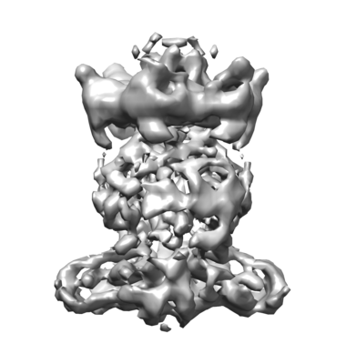 emd_8610.png emd_8610.png | 57.7 KB | ||
| Archive directory |  http://ftp.pdbj.org/pub/emdb/structures/EMD-8610ftp://ftp.pdbj.org/pub/emdb/structures/EMD-8610 http://ftp.pdbj.org/pub/emdb/structures/EMD-8610ftp://ftp.pdbj.org/pub/emdb/structures/EMD-8610 | HTTPS FTP |
-Related structure data
| Related structure data | 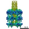 8608C 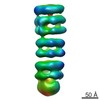 8611C 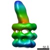 8612C  5uvnC 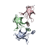 5uw2C 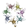 5uw8C 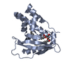 5uwaC 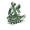 5uwbC C: citing same article ( |
|---|---|
| Similar structure data |
-Links
| EMDB pages | EMDB (EBI/PDBe) / EMDataResource |
|---|
-Map
| File | Download / File: emd_8610.map.gz / Format: CCP4 / Size: 1.2 MB / Type: IMAGE STORED AS FLOATING POINT NUMBER (4 BYTES) | ||||||||||||||||||||||||||||||||||||||||||||||||||||||||||||||||||||
|---|---|---|---|---|---|---|---|---|---|---|---|---|---|---|---|---|---|---|---|---|---|---|---|---|---|---|---|---|---|---|---|---|---|---|---|---|---|---|---|---|---|---|---|---|---|---|---|---|---|---|---|---|---|---|---|---|---|---|---|---|---|---|---|---|---|---|---|---|---|
| Annotation | E. coli MCE protein PqiB, periplasmic domain | ||||||||||||||||||||||||||||||||||||||||||||||||||||||||||||||||||||
| Projections & slices | Image control
Images are generated by Spider. | ||||||||||||||||||||||||||||||||||||||||||||||||||||||||||||||||||||
| Voxel size | X=Y=Z: 3.03 Å | ||||||||||||||||||||||||||||||||||||||||||||||||||||||||||||||||||||
| Density |
| ||||||||||||||||||||||||||||||||||||||||||||||||||||||||||||||||||||
| Symmetry | Space group: 1 | ||||||||||||||||||||||||||||||||||||||||||||||||||||||||||||||||||||
| Details | EMDB XML:
CCP4 map header:
| ||||||||||||||||||||||||||||||||||||||||||||||||||||||||||||||||||||
 Z (Sec.)
 Y (Row.)
 X (Col.)


-Supplemental data
- Sample components
-Entire : MlaFEDB
| Entire | Name: MlaFEDB |
|---|---|
| Components |
|
-Supramolecule #1: MlaFEDB
| Supramolecule | Name: MlaFEDB / type: complex / ID: 1 / Parent: 0 / Macromolecule list: all |
|---|---|
| Source (natural) | Organism: |
| Recombinant expression | Organism: |
-Macromolecule #1: MlaF
| Macromolecule | Name: MlaF / type: protein_or_peptide / ID: 1 / Enantiomer: LEVO |
|---|---|
| Source (natural) | Organism: |
| Recombinant expression | Organism: |
| Sequence | String: MEQSVANLVD MRDVSFTRGN RCIFDNISL TVPRGKITAI M GPSGIGKT TLLRLIGGQI AP DHGEILF DGENIPAMSR SRL YTVRKR MSMLFQSGAL FTDM NVFDN VAYPLREHTQ LPAPL LHST VMMKLEAVGL RGAAKL MPS ELSGGMARRA ALARAIA LE ...String: MEQSVANLVD MRDVSFTRGN RCIFDNISL TVPRGKITAI M GPSGIGKT TLLRLIGGQI AP DHGEILF DGENIPAMSR SRL YTVRKR MSMLFQSGAL FTDM NVFDN VAYPLREHTQ LPAPL LHST VMMKLEAVGL RGAAKL MPS ELSGGMARRA ALARAIA LE PDLIMFDEPF VGQDPITM G VLVKLISELN SALGVTCVV VSHDVPEVLS IADHAWILAD KKIVAHGSA QALQANPDPR V RQFLDGIA DGPVPFRYPA GD YHADLLP GS |
-Macromolecule #2: MlaE
| Macromolecule | Name: MlaE / type: protein_or_peptide / ID: 2 / Enantiomer: LEVO |
|---|---|
| Source (natural) | Organism: |
| Sequence | String: MLLNALASLG HKGIKTLRTF GRAGLMLFN ALVGKPEFRK H APLLVRQL YNVGVLSMLI IV VSGVFIG MVLGLQGYLV LTT YSAETS LGMLVALSLL RELG PVVAA LLFAGRAGSA LTAEI GLMR ATEQLSSMEM MAVDPL RRV ISPRFWAGVI SLPLLTV IF ...String: MLLNALASLG HKGIKTLRTF GRAGLMLFN ALVGKPEFRK H APLLVRQL YNVGVLSMLI IV VSGVFIG MVLGLQGYLV LTT YSAETS LGMLVALSLL RELG PVVAA LLFAGRAGSA LTAEI GLMR ATEQLSSMEM MAVDPL RRV ISPRFWAGVI SLPLLTV IF VAVGIWGGSL VGVSWKGI D SGFFWSAMQN AVDWRMDLV NCLIKSVVFA ITVTWISLFN GYDAIPTSA GISRATTRTV V HSSLAVLG LDFVLTALMF GN |
-Macromolecule #3: MlaD
| Macromolecule | Name: MlaD / type: protein_or_peptide / ID: 3 / Enantiomer: LEVO |
|---|---|
| Source (natural) | Organism: |
| Sequence | String: MHHHHHHQHQ HENLYFQGMQ TKKNEIWVG IFLLAALLAA L FVCLKAAN VTSIRTEPTY TL YATFDNI GGLKARSPVS IGG VVVGRV ADITLDPKTY LPRV TLEIE QRYNHIPDTS SLSIR TSGL LGEQYLALNV GFEDPE LGT AILKDGDTIQ DTKSAMV LE ...String: MHHHHHHQHQ HENLYFQGMQ TKKNEIWVG IFLLAALLAA L FVCLKAAN VTSIRTEPTY TL YATFDNI GGLKARSPVS IGG VVVGRV ADITLDPKTY LPRV TLEIE QRYNHIPDTS SLSIR TSGL LGEQYLALNV GFEDPE LGT AILKDGDTIQ DTKSAMV LE DLIGQFLYGS KGDDNKNS G DAPAAAPGNN ETTEPVGTT K |
-Macromolecule #4: MlaB
| Macromolecule | Name: MlaB / type: protein_or_peptide / ID: 4 / Enantiomer: LEVO |
|---|---|
| Source (natural) | Organism: |
| Sequence | String: MSESLSWMQT GDTLALSGEL DQDVLLPLW EMREEAVKGI T CIDLSRVS RVDTGGLALL LH LIDLAKK QGNNVTLQGV NDK VYTLAK LYNLPADVLP R |
-Experimental details
-Structure determination
| Method | cryo EM |
|---|---|
 Processing Processing | single particle reconstruction |
| Aggregation state | particle |
-Sample preparation
| Buffer | pH: 8 Details: 20 mM Tris pH 8.0 and 150 mM NaCl, and exchanged from DDM into amphipol A8-35 as described in the manuscript |
|---|---|
| Grid | Model: Quantifoil R1.2/1.3 / Material: COPPER / Mesh: 400 |
| Vitrification | Cryogen name: ETHANE / Instrument: FEI VITROBOT MARK III |
- Electron microscopy
| Microscope | FEI TITAN KRIOS |
|---|---|
| Image recording | Film or detector model: GATAN K2 SUMMIT (4k x 4k) / Average electron dose: 80.0 e/Å2 / Details: 80 e/A2 is total dose for 50 frames |
| Electron beam | Acceleration voltage: 300 kV / Electron source:  FIELD EMISSION GUN FIELD EMISSION GUN |
| Electron optics | Illumination mode: FLOOD BEAM / Imaging mode: BRIGHT FIELD |
| Experimental equipment |  Model: Titan Krios / Image courtesy: FEI Company |
+Image processing
-Atomic model buiding 1
| Details | crystal structure of MlaD and homology models for MlaF, MlaE and MlaB were fit into the density as described in the manuscript |
|---|---|
| Refinement | Space: REAL / Protocol: RIGID BODY FIT |
