 Movie
Movie Controller


+ Open data

- Basic information
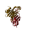
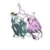
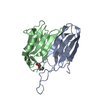
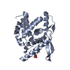
| Entry | Database: PDB / ID: 2ox5 | ||||||
|---|---|---|---|---|---|---|---|
| Title | The SoxYZ complex of Paracoccus pantotrophus | ||||||
 Components Components |
| ||||||
 Keywords Keywords | TRANSPORT PROTEIN / IMMUNOGLOBULIN-LIKE BETA-SANDWICH FOLD | ||||||
| Function / homology |  Function and homology information Function and homology informationSulphur oxidation protein SoxZ / Thiosulfate oxidation carrier complex protein SoxZ / Sulphur oxidation protein SoxZ / SoxY domain / Sulphur oxidation, SoxY / Ig-like SoxY domain / Ig-like SoxY domain superfamily / Sulfur oxidation protein SoxY / Immunoglobulin E-set / Immunoglobulin-like fold ...Sulphur oxidation protein SoxZ / Thiosulfate oxidation carrier complex protein SoxZ / Sulphur oxidation protein SoxZ / SoxY domain / Sulphur oxidation, SoxY / Ig-like SoxY domain / Ig-like SoxY domain superfamily / Sulfur oxidation protein SoxY / Immunoglobulin E-set / Immunoglobulin-like fold / Immunoglobulins / Immunoglobulin-like / Sandwich / Mainly Beta Similarity search - Domain/homology | ||||||
| Biological species |  Paracoccus denitrificans (bacteria) Paracoccus denitrificans (bacteria) | ||||||
| Method |  X-RAY DIFFRACTION / SYNCHROTRON / MAD / Resolution: 1.98 Å X-RAY DIFFRACTION / SYNCHROTRON / MAD / Resolution: 1.98 Å | ||||||
 Authors Authors | Bruno, S. / Sauve, V. / Berks, B.C. / Hemmings, A.M. | ||||||
 Citation Citation | Journal: J.Biol.Chem. / Year: 2007 Title: The SoxYZ Complex Carries Sulfur Cycle Intermediates on a Peptide Swinging Arm. Authors: Sauve, V. / Bruno, S. / Berks, B.C. / Hemmings, A.M. | ||||||
| History |
|
- Structure visualization
| Structure viewer | Molecule: MolmilJmol/JSmol |
|---|
- Downloads & links
-Download
| PDBx/mmCIF format | 2ox5.cif.gz | 191.9 KB | Display | PDBx/mmCIF format |
|---|---|---|---|---|
| PDB format | pdb2ox5.ent.gz | 151 KB | Display | PDB format |
| PDBx/mmJSON format | 2ox5.json.gz | Tree view | PDBx/mmJSON format | |
| Others |  Other downloads Other downloads |
-Validation report
| Arichive directory | https://data.pdbj.org/pub/pdb/validation_reports/ox/2ox5ftp://data.pdbj.org/pub/pdb/validation_reports/ox/2ox5 | HTTPS FTP |
|---|
-Related structure data


-Links
 PDBj
PDBj- Assembly
| Deposited unit | 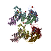
| ||||||||
|---|---|---|---|---|---|---|---|---|---|
| 1 | 
| ||||||||
| 2 | 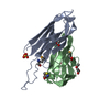
| ||||||||
| 3 | 
| ||||||||
| 4 | 
| ||||||||
| Unit cell |
| ||||||||
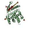
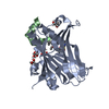
| Details | The biological assembly is a SoxYZ heterodimer. The crystallographic asymmetric unit contains two copies of this biological assembly. |
-Components
-Protein , 2 types, 8 molecules ZACEYBDF
| #1: Protein | Mass: 11781.041 Da / Num. of mol.: 4 Source method: isolated from a genetically manipulated source Source: (gene. exp.) Paracoccus denitrificans (bacteria) / Strain: LMD82.5T / Gene: soxZ / Plasmid: pVS005 / Production host: #2: Protein | Mass: 11436.581 Da / Num. of mol.: 4 Source method: isolated from a genetically manipulated source Source: (gene. exp.) Paracoccus denitrificans (bacteria) / Strain: LMD82.5T / Gene: soxY / Plasmid: pVS005 / Production host: |
|---|
-Non-polymers , 4 types, 907 molecules 


| #3: Chemical | ChemComp-ACT /  Mass: 59.044 Da / Num. of mol.: 8 / Source method: obtained synthetically / Formula: C2H3O2 Mass: 59.044 Da / Num. of mol.: 8 / Source method: obtained synthetically / Formula: C2H3O2#4: Chemical |  Mass: 96.063 Da / Num. of mol.: 3 / Source method: obtained synthetically / Formula: SO4 Mass: 96.063 Da / Num. of mol.: 3 / Source method: obtained synthetically / Formula: SO4#5: Chemical | ChemComp-EDO / |  Mass: 62.068 Da / Num. of mol.: 1 / Source method: obtained synthetically / Formula: C2H6O2 Mass: 62.068 Da / Num. of mol.: 1 / Source method: obtained synthetically / Formula: C2H6O2#6: Water | ChemComp-HOH / | Mass: 18.015 Da / Num. of mol.: 895 / Source method: isolated from a natural source / Formula: H2O |
|---|
-Details
| Has protein modification | Y |
|---|
-Experimental details
-Experiment
| Experiment | Method: X-RAY DIFFRACTION / Number of used crystals: 1 |
|---|
- Sample preparation
| Crystal | Density Matthews: 2.34 Å3/Da / Density % sol: 47.48 % |
|---|---|
| Crystal grow | Temperature: 293 K / Method: vapor diffusion, hanging drop / pH: 8 Details: 2 microl of protein at 10 mg/ml in 10 mM Tris-HCl pH 8.0, 5 mM beta-mercaptoethanol and 200 mM NaCl mixed with 2 microl of 28-30 % (w/v) PEG 3350, 100 mM sodium acetate pH 4.75, 200 mM ...Details: 2 microl of protein at 10 mg/ml in 10 mM Tris-HCl pH 8.0, 5 mM beta-mercaptoethanol and 200 mM NaCl mixed with 2 microl of 28-30 % (w/v) PEG 3350, 100 mM sodium acetate pH 4.75, 200 mM NH4SO4, VAPOR DIFFUSION, HANGING DROP, temperature 293K |
-Data collection
| Diffraction | Mean temperature: 100 K | ||||||||||||||||||||||||||||||||||||||||||||||||||||||||||||||||||||||||||||||||||||||||
|---|---|---|---|---|---|---|---|---|---|---|---|---|---|---|---|---|---|---|---|---|---|---|---|---|---|---|---|---|---|---|---|---|---|---|---|---|---|---|---|---|---|---|---|---|---|---|---|---|---|---|---|---|---|---|---|---|---|---|---|---|---|---|---|---|---|---|---|---|---|---|---|---|---|---|---|---|---|---|---|---|---|---|---|---|---|---|---|---|---|
| Diffraction source | Source: SYNCHROTRON / Site: ESRF  / Beamline: BM14 / Wavelength: 0.9793, 0.8731 / Beamline: BM14 / Wavelength: 0.9793, 0.8731 | ||||||||||||||||||||||||||||||||||||||||||||||||||||||||||||||||||||||||||||||||||||||||
| Detector | Type: MAR CCD 165 mm / Detector: CCD / Date: Apr 3, 2003 | ||||||||||||||||||||||||||||||||||||||||||||||||||||||||||||||||||||||||||||||||||||||||
| Radiation | Monochromator: Si(111) monochromator / Protocol: MAD / Monochromatic (M) / Laue (L): M / Scattering type: x-ray | ||||||||||||||||||||||||||||||||||||||||||||||||||||||||||||||||||||||||||||||||||||||||
| Radiation wavelength |
| ||||||||||||||||||||||||||||||||||||||||||||||||||||||||||||||||||||||||||||||||||||||||
| Reflection | Resolution: 1.981→102.147 Å / Num. all: 59443 / Num. obs: 59443 / % possible obs: 98.7 % / Observed criterion σ(F): 0 / Observed criterion σ(I): 0 / Redundancy: 4 % / Biso Wilson estimate: 19.7 Å2 / Rmerge(I) obs: 0.07 / Rsym value: 0.07 / Net I/σ(I): 8.6 | ||||||||||||||||||||||||||||||||||||||||||||||||||||||||||||||||||||||||||||||||||||||||
| Reflection shell |
|
- Processing
| Software |
| ||||||||||||||||||||||||||||||||||||||||||||||||||||||||||||||||||||||||||||||||||||||||||
|---|---|---|---|---|---|---|---|---|---|---|---|---|---|---|---|---|---|---|---|---|---|---|---|---|---|---|---|---|---|---|---|---|---|---|---|---|---|---|---|---|---|---|---|---|---|---|---|---|---|---|---|---|---|---|---|---|---|---|---|---|---|---|---|---|---|---|---|---|---|---|---|---|---|---|---|---|---|---|---|---|---|---|---|---|---|---|---|---|---|---|---|
| Refinement | Method to determine structure: MAD / Resolution: 1.98→50 Å / Cor.coef. Fo:Fc: 0.955 / Cor.coef. Fo:Fc free: 0.936 / SU B: 3.416 / SU ML: 0.099 / Isotropic thermal model: ISOTROPIC / Cross valid method: THROUGHOUT / σ(F): 0 / ESU R: 0.181 / ESU R Free: 0.153 / Stereochemistry target values: MAXIMUM LIKELIHOOD / Details: HYDROGENS HAVE BEEN ADDED IN THE RIDING POSITIONS
| ||||||||||||||||||||||||||||||||||||||||||||||||||||||||||||||||||||||||||||||||||||||||||
| Solvent computation | Ion probe radii: 0.8 Å / Shrinkage radii: 0.8 Å / VDW probe radii: 1.2 Å / Solvent model: MASK | ||||||||||||||||||||||||||||||||||||||||||||||||||||||||||||||||||||||||||||||||||||||||||
| Displacement parameters | Biso mean: 22.604 Å2
| ||||||||||||||||||||||||||||||||||||||||||||||||||||||||||||||||||||||||||||||||||||||||||
| Refinement step | Cycle: LAST / Resolution: 1.98→50 Å
| ||||||||||||||||||||||||||||||||||||||||||||||||||||||||||||||||||||||||||||||||||||||||||
| Refine LS restraints |
| ||||||||||||||||||||||||||||||||||||||||||||||||||||||||||||||||||||||||||||||||||||||||||
| LS refinement shell | Resolution: 1.981→2.032 Å / Total num. of bins used: 20
|
