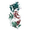
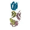
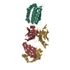
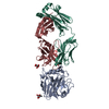
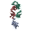
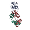
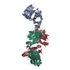
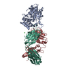
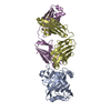
Entry Database : PDB / ID : 5ushTitle Structure of vaccinia virus D8 protein bound to human Fab vv66 Fab vv66 heavy chain Fab vv66 light chain IMV membrane protein Keywords / / / / / / Function / homology Function Domain/homology Component
/ / / / / / / / / / / / / / / / / / / / / / Biological species Homo sapiens (human)Method / / / / Resolution : 2.3 Å Authors Zajonc, D.M. Funding support Organization Grant number Country National Institutes of Health/National Institute of General Medical Sciences (NIH/NIGMS) HHSN272200900048C
Journal : J. Biol. Chem. / Year : 2018Title : Structure-function characterization of three human antibodies targeting the vaccinia virus adhesion molecule D8.Authors: Matho, M.H. / Schlossman, A. / Gilchuk, I.M. / Miller, G. / Mikulski, Z. / Hupfer, M. / Wang, J. / Bitra, A. / Meng, X. / Xiang, Y. / Kaever, T. / Doukov, T. / Ley, K. / Crotty, S. / Peters, ... Authors : Matho, M.H. / Schlossman, A. / Gilchuk, I.M. / Miller, G. / Mikulski, Z. / Hupfer, M. / Wang, J. / Bitra, A. / Meng, X. / Xiang, Y. / Kaever, T. / Doukov, T. / Ley, K. / Crotty, S. / Peters, B. / Hsieh-Wilson, L.C. / Crowe, J.E. / Zajonc, D.M. History Deposition Feb 13, 2017 Deposition site / Processing site Revision 1.0 Sep 20, 2017 Provider / Type Revision 1.1 Nov 22, 2017 Group / Category / citation_authorItem _citation.country / _citation.journal_abbrev ... _citation.country / _citation.journal_abbrev / _citation.journal_id_ASTM / _citation.journal_id_CSD / _citation.journal_id_ISSN / _citation.pdbx_database_id_DOI / _citation.pdbx_database_id_PubMed / _citation.title / _citation.year Revision 1.2 Jan 17, 2018 Group / Category Item _citation.journal_volume / _citation.page_first ... _citation.journal_volume / _citation.page_first / _citation.page_last / _citation.year Revision 1.3 Dec 4, 2019 Group / Category / Item Revision 1.4 Oct 4, 2023 Group / Database references / Refinement descriptionCategory chem_comp_atom / chem_comp_bond ... chem_comp_atom / chem_comp_bond / database_2 / pdbx_initial_refinement_model Item / _database_2.pdbx_database_accessionRevision 1.5 Nov 6, 2024 Group / Category / pdbx_modification_feature
Show all Show less
 Movie
Movie Controller


 Open data
 Basic information
 Components
 Keywords
 Function and homology information
 Vaccinia virus
 Homo sapiens (human)
 X-RAY DIFFRACTION /
  Authors
 United States, 1items
  Citation
 Structure visualization
 Downloads & links
 Other downloads


 PDBj
PDBj

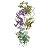
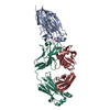
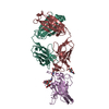
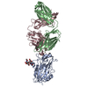
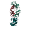
 Assembly
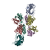

 Mass: 18.015 Da / Num. of mol.: 174 / Source method: isolated from a natural source / Formula: H2O
 Sample preparation
 Processing