[English] 日本語
Yorodumi
- PDB-5ur2: Crystal structure of proline utilization A (PutA) from Bdellovibr... -

+
Open data


ID or keywords:

Loading...

-
Basic information

Entry
Database: PDB / ID: 5ur2
TitleCrystal structure of proline utilization A (PutA) from Bdellovibrio bacteriovorus inactivated by N-propargylglycine
ComponentsBifunctional protein PutA
KeywordsOXIDOREDUCTASE / FLAVOENZYME / ROSSMANN FOLD / ALDEHYDE DEHYDROGENASE / FLAVIN ADENINE DINUCLEOTIDE / NICOTINAMIDE ADENINE DINUCLEOTIDE / PROLINE CATABOLISM / SUBSTRATE CHANNELING / BIFUNCTIONAL ENZYME / MECHANISM-BASED INACTIVATION
Function / homology
Function and homology information


proline dehydrogenase / proline dehydrogenase activity / L-glutamate gamma-semialdehyde dehydrogenase / 1-pyrroline-5-carboxylate dehydrogenase activity / proline catabolic process to glutamate / proline biosynthetic process / DNA-binding transcription factor activity / DNA binding
Similarity search - Function
Proline utilization A, N-terminal / Proline utilization A N-terminal domain / 1-pyrroline-5-carboxylate dehydrogenase / Bifunctional protein PutA / Proline dehydrogenase domain / Proline dehydrogenase / FAD-linked oxidoreductase-like / Aldehyde dehydrogenase, glutamic acid active site / Aldehyde dehydrogenases glutamic acid active site. / Aldehyde dehydrogenase, cysteine active site ...Proline utilization A, N-terminal / Proline utilization A N-terminal domain / 1-pyrroline-5-carboxylate dehydrogenase / Bifunctional protein PutA / Proline dehydrogenase domain / Proline dehydrogenase / FAD-linked oxidoreductase-like / Aldehyde dehydrogenase, glutamic acid active site / Aldehyde dehydrogenases glutamic acid active site. / Aldehyde dehydrogenase, cysteine active site / Aldehyde dehydrogenases cysteine active site. / Aldehyde dehydrogenase domain / Aldehyde dehydrogenase family / Aldehyde dehydrogenase, N-terminal / Aldehyde dehydrogenase, C-terminal / Aldehyde/histidinol dehydrogenase
Similarity search - Domain/homology
Chem-P5F / Bifunctional protein PutA
Similarity search - Component
Biological speciesBdellovibrio bacteriovorus (bacteria)
MethodX-RAY DIFFRACTION / SYNCHROTRON / MOLECULAR REPLACEMENT / Resolution: 2.23 Å
AuthorsTanner, J.J.
Funding support United States, 1items
OrganizationGrant numberCountry
National Institutes of Health/National Institute of General Medical Sciences (NIH/NIGMS)R01GM065546 United States
Citation
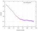
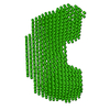
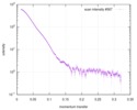
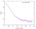
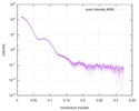
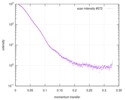
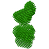
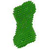
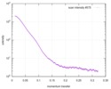
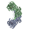
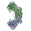
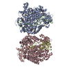
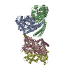
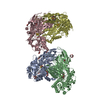
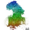
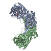
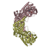
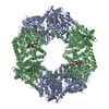
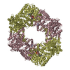
Journal: FEBS J / Year: 2017
Title: Biophysical investigation of type A PutAs reveals a conserved core oligomeric structure.
Authors: David A Korasick / Harkewal Singh / Travis A Pemberton / Min Luo / Richa Dhatwalia / John J Tanner /
Abstract: Many enzymes form homooligomers, yet the functional significance of self-association is seldom obvious. Herein, we examine the connection between oligomerization and catalytic function for proline ...Many enzymes form homooligomers, yet the functional significance of self-association is seldom obvious. Herein, we examine the connection between oligomerization and catalytic function for proline utilization A (PutA) enzymes. PutAs are bifunctional enzymes that catalyze both reactions of proline catabolism. Type A PutAs are the smallest members of the family, possessing a minimal domain architecture consisting of N-terminal proline dehydrogenase and C-terminal l-glutamate-γ-semialdehyde dehydrogenase modules. Type A PutAs form domain-swapped dimers, and in one case (Bradyrhizobium japonicum PutA), two of the dimers assemble into a ring-shaped tetramer. Whereas the dimer has a clear role in substrate channeling, the functional significance of the tetramer is unknown. To address this question, we performed structural studies of four-type A PutAs from two clades of the PutA tree. The crystal structure of Bdellovibrio bacteriovorus PutA covalently inactivated by N-propargylglycine revealed a fold and substrate-channeling tunnel similar to other PutAs. Small-angle X-ray scattering (SAXS) and analytical ultracentrifugation indicated that Bdellovibrio PutA is dimeric in solution, in contrast to the prediction from crystal packing of a stable tetrameric assembly. SAXS studies of two other type A PutAs from separate clades also suggested that the dimer predominates in solution. To assess whether the tetramer of B. japonicum PutA is necessary for catalytic function, a hot spot disruption mutant that cleanly produces dimeric protein was generated. The dimeric variant exhibited kinetic parameters similar to the wild-type enzyme. These results implicate the domain-swapped dimer as the core structural and functional unit of type A PutAs.
ENZYMES: Proline dehydrogenase (EC 1.5.5.2); l-glutamate-γ-semialdehyde dehydrogenase (EC 1.2.1.88).
DATABASES: The atomic coordinates and structure factor amplitudes have been deposited in the Protein Data Bank under accession number 5UR2. The SAXS data have been deposited in the SASBDB under the ...DATABASES: The atomic coordinates and structure factor amplitudes have been deposited in the Protein Data Bank under accession number 5UR2. The SAXS data have been deposited in the SASBDB under the following accession codes: SASDCP3 (BbPutA), SASDCQ3 (DvPutA 1.5 mg·mL ), SASDCX3 (DvPutA 3.0 mg·mL ), SASDCY3 (DvPutA 4.5 mg·mL ), SASDCR3 (LpPutA 3.0 mg·mL ), SASDCV3 (LpPutA 5.0 mg·mL ), SASDCW3 (LpPutA 8.0 mg·mL ), SASDCS3 (BjPutA 2.3 mg·mL ), SASDCT3 (BjPutA 4.7 mg·mL ), SASDCU3 (BjPutA 7.0 mg·mL ), SASDCZ3 (R51E 2.3 mg·mL ), SASDC24 (R51E 4.7 mg·mL ), SASDC34 (R51E 7.0 mg·mL ).
History
DepositionFeb 9, 2017Deposition site: RCSB / Processing site: RCSB
Revision 1.0Aug 2, 2017Provider: repository / Type: Initial release
Revision 1.1Oct 4, 2017Group: Advisory / Database references / Category: citation / pdbx_unobs_or_zero_occ_atoms
Item: _citation.journal_volume / _citation.page_first / _citation.page_last
Revision 1.2Jan 1, 2020Group: Author supporting evidence / Category: pdbx_audit_support / Item: _pdbx_audit_support.funding_organization
Revision 1.3Oct 4, 2023Group: Data collection / Database references / Refinement description
Category: chem_comp_atom / chem_comp_bond ...chem_comp_atom / chem_comp_bond / database_2 / pdbx_initial_refinement_model
Item: _database_2.pdbx_DOI / _database_2.pdbx_database_accession

-
Structure visualization

Structure viewerMolecule:
MolmilJmol/JSmol

Downloads & links

-
Assembly

Deposited unit
A: Bifunctional protein PutA
B: Bifunctional protein PutA
C: Bifunctional protein PutA
D: Bifunctional protein PutA
hetero molecules


Theoretical massNumber of molelcules
Total (without water)442,1478
Polymers438,5524
Non-polymers3,5954
Water18,8801048
1
A: Bifunctional protein PutA
B: Bifunctional protein PutA
hetero molecules


Theoretical massNumber of molelcules
Total (without water)221,0734
Polymers219,2762
Non-polymers1,7972
Water362
TypeNameSymmetry operationNumber
identity operation1_555x,y,z1
Buried area7770 Å2
ΔGint-33 kcal/mol
Surface area66390 Å2
MethodPISA
2
C: Bifunctional protein PutA
D: Bifunctional protein PutA
hetero molecules


Theoretical massNumber of molelcules
Total (without water)221,0734
Polymers219,2762
Non-polymers1,7972
Water362
TypeNameSymmetry operationNumber
identity operation1_555x,y,z1
Buried area7750 Å2
ΔGint-33 kcal/mol
Surface area66390 Å2
MethodPISA
3
A: Bifunctional protein PutA
B: Bifunctional protein PutA
hetero molecules

A: Bifunctional protein PutA
B: Bifunctional protein PutA
hetero molecules


Theoretical massNumber of molelcules
Total (without water)442,1478
Polymers438,5524
Non-polymers3,5954
Water724
TypeNameSymmetry operationNumber
identity operation1_555x,y,z1
crystal symmetry operation2_555-x,y,-z1
Buried area20030 Å2
ΔGint-85 kcal/mol
Surface area128280 Å2
MethodPISA
4
C: Bifunctional protein PutA
D: Bifunctional protein PutA
hetero molecules

C: Bifunctional protein PutA
D: Bifunctional protein PutA
hetero molecules


Theoretical massNumber of molelcules
Total (without water)442,1478
Polymers438,5524
Non-polymers3,5954
Water724
TypeNameSymmetry operationNumber
identity operation1_555x,y,z1
crystal symmetry operation2_555-x,y,-z1
Buried area19800 Å2
ΔGint-92 kcal/mol
Surface area128470 Å2
MethodPISA
Unit cell
Length a, b, c (Å)144.330, 158.649, 221.152
Angle α, β, γ (deg.)90.00, 90.00, 90.00
Int Tables number18
Space group name H-MP21221
Noncrystallographic symmetry (NCS)NCS domain:
IDEns-ID
11
21
31
41
12
22
32
42

NCS domain segments:
Dom-IDComponent-IDEns-IDSelection details
111CHAIN A AND RESID 1001
211CHAIN B AND RESID 1001
311CHAIN C AND RESID 1001
411CHAIN D AND RESID 1001
112(CHAIN A AND ((RESID 3 AND (NAME O OR NAME...
212(CHAIN B AND ((RESID 3 AND (NAME O OR NAME...
312(CHAIN C AND ((RESID 3 AND (NAME N OR NAME...
412(CHAIN D AND ((RESID 3 AND (NAME O OR NAME...

NCS ensembles :
ID
1
2

-
Components

#1: Protein
Bifunctional protein PutA


Mass: 109638.047 Da / Num. of mol.: 4
Source method: isolated from a genetically manipulated source
Source: (gene. exp.) Bdellovibrio bacteriovorus (strain ATCC 15356 / DSM 50701 / NCIB 9529 / HD100) (bacteria)
Strain: ATCC 15356 / DSM 50701 / NCIB 9529 / HD100 / Gene: putA, Bd1251 / Production host: Escherichia coli (E. coli) / References: UniProt: Q6MNK1
#2: Chemical
ChemComp-P5F / N-propargylglycine-modified flavin adenine dinucleotide


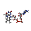
Mass: 898.664 Da / Num. of mol.: 4 / Source method: obtained synthetically / Formula: C32H40N10O17P2
#3: Water ChemComp-HOH / water


Mass: 18.015 Da / Num. of mol.: 1048 / Source method: isolated from a natural source / Formula: H2O

-
Experimental details

-
Experiment

ExperimentMethod: X-RAY DIFFRACTION / Number of used crystals: 1

-
Sample preparation

CrystalDensity Matthews: 2.89 Å3/Da / Density % sol: 57.39 %
Crystal growTemperature: 293 K / Method: microbatch / pH: 7.5 / Details: 19% PEG 3350, 0.25M KSCN

-
Data collection

DiffractionMean temperature: 100 K
Diffraction sourceSource: SYNCHROTRON / Site: APS / Beamline: 24-ID-E / Wavelength: 0.9792 Å
DetectorType: ADSC QUANTUM 315r / Detector: CCD / Date: Jul 1, 2012
RadiationProtocol: SINGLE WAVELENGTH / Monochromatic (M) / Laue (L): M / Scattering type: x-ray
Radiation wavelengthWavelength: 0.9792 Å / Relative weight: 1
ReflectionResolution: 2.228→128.909 Å / Num. obs: 241211 / % possible obs: 97.9 % / Redundancy: 3.9 % / Biso Wilson estimate: 27.58 Å2 / Rsym value: 0.095 / Net I/σ(I): 10
Reflection shellResolution: 2.23→2.35 Å / Redundancy: 3.9 % / Rmerge(I) obs: 0.493 / Mean I/σ(I) obs: 1.6 / Rsym value: 0.493 / % possible all: 98.3

-
Processing

Software
NameVersionClassification
PHENIX1.10.1_2155refinement
SCALA3.3.16data scaling
PDB_EXTRACT3.22data extraction
XDSdata reduction
MOLREPphasing
RefinementMethod to determine structure: MOLECULAR REPLACEMENT
Starting model: 4NME
Resolution: 2.23→128.91 Å / SU ML: 0.27 / Cross valid method: FREE R-VALUE / σ(F): 0 / Phase error: 22.43 / Stereochemistry target values: ML
RfactorNum. reflection% reflection
Rfree0.228 12095 5.02 %
Rwork0.183 --
obs0.186 241137 97.6 %
Solvent computationShrinkage radii: 0.9 Å / VDW probe radii: 1.11 Å / Solvent model: FLAT BULK SOLVENT MODEL
Displacement parametersBiso mean: 31.34 Å2
Refinement stepCycle: LAST / Resolution: 2.23→128.91 Å
ProteinNucleic acidLigandSolventTotal
Num. atoms29556 0 224 1048 30828
Refine LS restraints
Refine-IDTypeDev idealNumber
X-RAY DIFFRACTIONf_bond_d0.00830428
X-RAY DIFFRACTIONf_angle_d0.87241246
X-RAY DIFFRACTIONf_dihedral_angle_d12.24918440
X-RAY DIFFRACTIONf_chiral_restr0.0524564
X-RAY DIFFRACTIONf_plane_restr0.0055347
Refine LS restraints NCS
Ens-IDDom-IDAuth asym-IDNumberRefine-IDType
11A0X-RAY DIFFRACTIONPOSITIONAL
12B0X-RAY DIFFRACTIONPOSITIONAL
13C0X-RAY DIFFRACTIONPOSITIONAL
14D0X-RAY DIFFRACTIONPOSITIONAL
21A17801X-RAY DIFFRACTIONPOSITIONAL
22B17801X-RAY DIFFRACTIONPOSITIONAL
23C17801X-RAY DIFFRACTIONPOSITIONAL
24D17801X-RAY DIFFRACTIONPOSITIONAL
LS refinement shell
Resolution (Å)Rfactor RfreeNum. reflection RfreeRfactor RworkNum. reflection RworkRefine-ID% reflection obs (%)
2.2276-2.25290.31344170.24837231X-RAY DIFFRACTION94
2.2529-2.27940.29524170.23597730X-RAY DIFFRACTION100
2.2794-2.30720.28154240.21827697X-RAY DIFFRACTION100
2.3072-2.33640.27284030.21747717X-RAY DIFFRACTION99
2.3364-2.36710.29814160.22357709X-RAY DIFFRACTION99
2.3671-2.39960.27093350.21727772X-RAY DIFFRACTION100
2.3996-2.43390.28514100.21657691X-RAY DIFFRACTION99
2.4339-2.47020.28294000.21627711X-RAY DIFFRACTION99
2.4702-2.50880.28923900.21487732X-RAY DIFFRACTION99
2.5088-2.54990.2794180.20727690X-RAY DIFFRACTION99
2.5499-2.59390.26884500.20197699X-RAY DIFFRACTION99
2.5939-2.64110.27544170.27653X-RAY DIFFRACTION99
2.6411-2.69190.24044100.19617634X-RAY DIFFRACTION98
2.6919-2.74680.24443880.19767623X-RAY DIFFRACTION98
2.7468-2.80660.27883680.20237150X-RAY DIFFRACTION92
2.8066-2.87180.24623670.2127321X-RAY DIFFRACTION94
2.8718-2.94370.2634250.21217694X-RAY DIFFRACTION99
2.9437-3.02330.27524040.21577707X-RAY DIFFRACTION99
3.0233-3.11220.26584020.21677763X-RAY DIFFRACTION99
3.1122-3.21270.25043630.20537732X-RAY DIFFRACTION99
3.2127-3.32750.2444060.20357725X-RAY DIFFRACTION99
3.3275-3.46080.24553810.19327763X-RAY DIFFRACTION99
3.4608-3.61830.2274330.18687682X-RAY DIFFRACTION98
3.6183-3.80910.22254360.17787597X-RAY DIFFRACTION98
3.8091-4.04770.18174000.15417515X-RAY DIFFRACTION95
4.0477-4.36030.17363790.14037130X-RAY DIFFRACTION91
4.3603-4.79910.16314110.12967788X-RAY DIFFRACTION98
4.7991-5.49350.15894170.13767791X-RAY DIFFRACTION98
5.4935-6.92120.17824100.15137709X-RAY DIFFRACTION96
6.9212-129.12570.1723980.14757686X-RAY DIFFRACTION93
Refinement TLS params.

Method: refined / Refine-ID: X-RAY DIFFRACTION

IDL112)L122)L132)L222)L232)L332)S11 (Å °)S12 (Å °)S13 (Å °)S21 (Å °)S22 (Å °)S23 (Å °)S31 (Å °)S32 (Å °)S33 (Å °)T112)T122)T132)T222)T232)T332)Origin x (Å)Origin y (Å)Origin z (Å)
10.4580.2146-0.38830.2131-0.15240.6917-0.0326-0.0057-0.0255-0.0068-0.0109-0.020.0476-0.10260.04470.13910.0084-0.02020.1926-0.02510.2001-44.530537.9127-4.5579
20.85290.1515-0.04630.3417-0.0070.2334-0.04680.09090.1844-0.03440.0315-0.0189-0.07080.04980.01890.20060.0073-0.02850.17330.00160.20485.539455.539-38.2223
30.1868-0.02240.070.2875-0.32120.97520.0209-0.0519-0.0621-0.01180.05040.04070.0947-0.1048-0.07460.2206-0.01170.03690.17770.00810.2123-34.8576-36.837716.4998
40.4650.2036-0.2470.4164-0.19620.6114-0.03390.0982-0.01340.01390.0546-0.05250.0436-0.0408-0.02130.1820.00610.00150.1639-0.03060.1795-17.9129-20.1552-41.771
Refinement TLS group
IDRefine-IDRefine TLS-IDSelection details
1X-RAY DIFFRACTION1CHAIN A
2X-RAY DIFFRACTION2CHAIN B
3X-RAY DIFFRACTION3CHAIN C
4X-RAY DIFFRACTION4CHAIN D

+
About Yorodumi

-
News

-
Feb 9, 2022. New format data for meta-information of EMDB entries

New format data for meta-information of EMDB entries

  • Version 3 of the EMDB header file is now the official format.
  • The previous official version 1.9 will be removed from the archive.

Related info.:EMDB header

External links:wwPDB to switch to version 3 of the EMDB data model

-
Aug 12, 2020. Covid-19 info

Covid-19 info

URL: https://pdbjlvh1.pdbj.org/emnavi/covid19.php

New page: Covid-19 featured information page in EM Navigator.

Related info.:Covid-19 info / Mar 5, 2020. Novel coronavirus structure data

+
Mar 5, 2020. Novel coronavirus structure data

Novel coronavirus structure data

Related info.:Yorodumi Speices / Aug 12, 2020. Covid-19 info

External links:COVID-19 featured content - PDBj / Molecule of the Month (242):Coronavirus Proteases

+
Jan 31, 2019. EMDB accession codes are about to change! (news from PDBe EMDB page)

EMDB accession codes are about to change! (news from PDBe EMDB page)

  • The allocation of 4 digits for EMDB accession codes will soon come to an end. Whilst these codes will remain in use, new EMDB accession codes will include an additional digit and will expand incrementally as the available range of codes is exhausted. The current 4-digit format prefixed with “EMD-” (i.e. EMD-XXXX) will advance to a 5-digit format (i.e. EMD-XXXXX), and so on. It is currently estimated that the 4-digit codes will be depleted around Spring 2019, at which point the 5-digit format will come into force.
  • The EM Navigator/Yorodumi systems omit the EMD- prefix.

Related info.:Q: What is EMD? / ID/Accession-code notation in Yorodumi/EM Navigator

External links:EMDB Accession Codes are Changing Soon! / Contact to PDBj

+
Jul 12, 2017. Major update of PDB

Major update of PDB

  • wwPDB released updated PDB data conforming to the new PDBx/mmCIF dictionary.
  • This is a major update changing the version number from 4 to 5, and with Remediation, in which all the entries are updated.
  • In this update, many items about electron microscopy experimental information are reorganized (e.g. em_software).
  • Now, EM Navigator and Yorodumi are based on the updated data.

External links:wwPDB Remediation / Enriched Model Files Conforming to OneDep Data Standards Now Available in the PDB FTP Archive

-
Yorodumi

Thousand views of thousand structures

  • Yorodumi is a browser for structure data from EMDB, PDB, SASBDB, etc.
  • This page is also the successor to EM Navigator detail page, and also detail information page/front-end page for Omokage search.
  • The word "yorodu" (or yorozu) is an old Japanese word meaning "ten thousand". "mi" (miru) is to see.

Related info.:EMDB / PDB / SASBDB / Comparison of 3 databanks / Yorodumi Search / Aug 31, 2016. New EM Navigator & Yorodumi / Yorodumi Papers / Jmol/JSmol / Function and homology information / Changes in new EM Navigator and Yorodumi

Read more