 Movie
Movie Controller

[English] 日本語
 Yorodumi
- PDB-5njt: Structure of the Bacillus subtilis hibernating 100S ribosome reve... -
+ Open data

- Basic information
| Entry | Database: PDB / ID: 5njt | ||||||
|---|---|---|---|---|---|---|---|
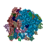
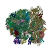
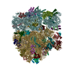
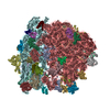
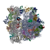
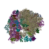
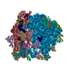
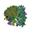
| Title | Structure of the Bacillus subtilis hibernating 100S ribosome reveals the basis for 70S dimerization. | ||||||
 Components Components |
| ||||||
 Keywords Keywords | TRANSLATION / 100S / Bacillus subtilis / cryo-EM / Hibernation / HPF / RMF / rRNA / YvyD | ||||||
| Function / homology |  Function and homology information Function and homology informationnegative regulation of translational elongation / positive regulation of rRNA processing / nucleoid / ribosomal small subunit binding / rRNA processing / large ribosomal subunit / transferase activity / ribosome biogenesis / ribosomal small subunit biogenesis / 5S rRNA binding ...negative regulation of translational elongation / positive regulation of rRNA processing / nucleoid / ribosomal small subunit binding / rRNA processing / large ribosomal subunit / transferase activity / ribosome biogenesis / ribosomal small subunit biogenesis / 5S rRNA binding / ribosomal large subunit assembly / small ribosomal subunit / small ribosomal subunit rRNA binding / large ribosomal subunit rRNA binding / cytosolic small ribosomal subunit / cytosolic large ribosomal subunit / cytoplasmic translation / tRNA binding / negative regulation of translation / rRNA binding / structural constituent of ribosome / ribosome / translation / ribonucleoprotein complex / response to antibiotic / mRNA binding / DNA binding / RNA binding / zinc ion binding / metal ion binding / cytosol / cytoplasm Similarity search - Function | ||||||
| Biological species |  | ||||||
| Method | ELECTRON MICROSCOPY / single particle reconstruction / cryo EM / Resolution: 3.8 Å | ||||||
 Authors Authors | Beckert, B. / Abdelshahid, M. / Schaefer, H. / Steinchen, W. / Arenz, S. / Berninghausen, O. / Beckmann, R. / Bange, G. / Turgay, K. / Wilson, D.N. | ||||||
| Funding support |  Germany, 1items Germany, 1items
| ||||||
 Citation Citation | Journal: EMBO J / Year: 2017 Title: Structure of the hibernating 100S ribosome reveals the basis for 70S dimerization. Authors: Bertrand Beckert / Maha Abdelshahid / Heinrich Schäfer / Wieland Steinchen / Stefan Arenz / Otto Berninghausen / Roland Beckmann / Gert Bange / Kürşad Turgay / Daniel N Wilson / Abstract: Under stress conditions, such as nutrient deprivation, bacteria enter into a hibernation stage, which is characterized by the appearance of 100S ribosomal particles. In , dimerization of 70S ...Under stress conditions, such as nutrient deprivation, bacteria enter into a hibernation stage, which is characterized by the appearance of 100S ribosomal particles. In , dimerization of 70S ribosomes into 100S requires the action of the ribosome modulation factor (RMF) and the hibernation-promoting factor (HPF). Most other bacteria lack RMF and instead contain a long form HPF (LHPF), which is necessary and sufficient for 100S formation. While some structural information exists as to how RMF and HPF mediate formation of 100S (100S), structural insight into 100S formation by LHPF has so far been lacking. Here we present a cryo-EM structure of the hibernating 100S (100S), revealing that the C-terminal domain (CTD) of the LHPF occupies a site on the 30S platform distinct from RMF Moreover, unlike RMF, the HPF-CTD is directly involved in forming the dimer interface, thereby illustrating the divergent mechanisms by which 100S formation is mediated in the majority of bacteria that contain LHPF, compared to some γ-proteobacteria, such as . | ||||||
| History |
|
- Structure visualization
| Movie |
Movie viewer |
|---|---|
| Structure viewer | Molecule: MolmilJmol/JSmol |
- Downloads & links
-Download
| PDBx/mmCIF format | 5njt.cif.gz | 3 MB | Display | PDBx/mmCIF format |
|---|---|---|---|---|
| PDB format | pdb5njt.ent.gz | Display | PDB format | |
| PDBx/mmJSON format | 5njt.json.gz | Tree view | PDBx/mmJSON format | |
| Others |  Other downloads Other downloads |
-Validation report
| Arichive directory | https://data.pdbj.org/pub/pdb/validation_reports/nj/5njtftp://data.pdbj.org/pub/pdb/validation_reports/nj/5njt | HTTPS FTP |
|---|
-Related structure data
| Related structure data | 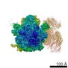 3656MC 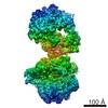 3664C M: map data used to model this data C: citing same article ( |
|---|---|
| Similar structure data |
-Links
 PDBj
PDBj


- Assembly
| Deposited unit | 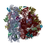
|
|---|---|
| 1 |
|
-Components
-RNA chain , 3 types, 3 molecules AUV
| #1: RNA chain | Mass: 500263.219 Da / Num. of mol.: 1 / Source method: isolated from a natural source Source: (natural) Plasmid details: Bacillus Genetic Stock Center, Columbus, Ohio, USA -1A1 Variant: trpC2 / References: GenBank: 225184640 |
|---|---|
| #21: RNA chain | Mass: 947975.188 Da / Num. of mol.: 1 / Source method: isolated from a natural source Source: (natural) References: GenBank: 467326 |
| #22: RNA chain | Mass: 36157.520 Da / Num. of mol.: 1 / Source method: isolated from a natural source Source: (natural) References: GenBank: 728882887 |
-30S ribosomal protein ... , 19 types, 19 molecules BCDEFGHIJKLMNOPQRST
| #2: Protein | Mass: 25730.895 Da / Num. of mol.: 1 / Source method: isolated from a natural source Source: (natural) References: UniProt: P21464 |
|---|---|
| #3: Protein | Mass: 23488.898 Da / Num. of mol.: 1 / Source method: isolated from a natural source Source: (natural) References: UniProt: P21465 |
| #4: Protein | Mass: 22743.074 Da / Num. of mol.: 1 / Source method: isolated from a natural source Source: (natural) References: UniProt: P21466 |
| #5: Protein | Mass: 17519.430 Da / Num. of mol.: 1 / Source method: isolated from a natural source Source: (natural) References: UniProt: P21467 |
| #6: Protein | Mass: 11140.548 Da / Num. of mol.: 1 / Source method: isolated from a natural source Source: (natural) References: UniProt: P21468 |
| #7: Protein | Mass: 17530.369 Da / Num. of mol.: 1 / Source method: isolated from a natural source Source: (natural) References: UniProt: P21469 |
| #8: Protein | Mass: 14770.229 Da / Num. of mol.: 1 / Source method: isolated from a natural source Source: (natural) References: UniProt: P12879 |
| #9: Protein | Mass: 14335.504 Da / Num. of mol.: 1 / Source method: isolated from a natural source Source: (natural) References: UniProt: P21470 |
| #10: Protein | Mass: 11687.661 Da / Num. of mol.: 1 / Source method: isolated from a natural source Source: (natural) References: UniProt: P21471 |
| #11: Protein | Mass: 12476.212 Da / Num. of mol.: 1 / Source method: isolated from a natural source Source: (natural) References: UniProt: P04969 |
| #12: Protein | Mass: 15117.538 Da / Num. of mol.: 1 / Source method: isolated from a natural source Source: (natural) References: UniProt: P21472 |
| #13: Protein | Mass: 12599.545 Da / Num. of mol.: 1 / Source method: isolated from a natural source Source: (natural) References: UniProt: P20282 |
| #14: Protein | Mass: 7132.607 Da / Num. of mol.: 1 / Source method: isolated from a natural source Source: (natural) References: UniProt: P12878 |
| #15: Protein | Mass: 10466.028 Da / Num. of mol.: 1 / Source method: isolated from a natural source Source: (natural) References: UniProt: P21473 |
| #16: Protein | Mass: 10022.637 Da / Num. of mol.: 1 / Source method: isolated from a natural source Source: (natural) References: UniProt: P21474 |
| #17: Protein | Mass: 10089.784 Da / Num. of mol.: 1 / Source method: isolated from a natural source Source: (natural) References: UniProt: P12874 |
| #18: Protein | Mass: 8245.746 Da / Num. of mol.: 1 / Source method: isolated from a natural source Source: (natural) References: UniProt: P21475 |
| #19: Protein | Mass: 9185.659 Da / Num. of mol.: 1 / Source method: isolated from a natural source Source: (natural) References: UniProt: P21476 |
| #20: Protein | Mass: 9393.906 Da / Num. of mol.: 1 / Source method: isolated from a natural source Source: (natural) References: UniProt: P21477 |
+50S ribosomal protein ... , 28 types, 28 molecules WXYZabcdefghijklmnopqrstuvwy
-Protein , 1 types, 1 molecules x
| #51: Protein | Mass: 12355.955 Da / Num. of mol.: 1 / Source method: isolated from a natural source Source: (natural) References: UniProt: P28368 |
|---|
-Experimental details
-Experiment
| Experiment | Method: ELECTRON MICROSCOPY |
|---|---|
| EM experiment | Aggregation state: PARTICLE / 3D reconstruction method: single particle reconstruction |
- Sample preparation
| Component | Name: Structure of the Bacillus subtilis hibernating 100S ribosome reveals the basis for 70S dimerization Type: RIBOSOME / Entity ID: all / Source: NATURAL | ||||||||||||||||||||
|---|---|---|---|---|---|---|---|---|---|---|---|---|---|---|---|---|---|---|---|---|---|
| Source (natural) | Organism: | ||||||||||||||||||||
| Buffer solution | pH: 7.5 | ||||||||||||||||||||
| Buffer component |
| ||||||||||||||||||||
| Specimen | Embedding applied: NO / Shadowing applied: NO / Staining applied: NO / Vitrification applied: YES Details: 4 OD260/ml ml Bs100S sample were applied to 2 nm pre-coated Quantifoil R3/3 holey carbon supported grids and vitrified using Vitrobot Mark IV (FEI Company) | ||||||||||||||||||||
| Specimen support | Grid type: Quantifoil R3/3 | ||||||||||||||||||||
| Vitrification | Cryogen name: ETHANE |
- Electron microscopy imaging
| Experimental equipment |  Model: Titan Krios / Image courtesy: FEI Company |
|---|---|
| Microscopy | Model: FEI TITAN KRIOS |
| Electron gun | Electron source:  FIELD EMISSION GUN / Accelerating voltage: 300 kV / Illumination mode: FLOOD BEAM FIELD EMISSION GUN / Accelerating voltage: 300 kV / Illumination mode: FLOOD BEAM |
| Electron lens | Mode: BRIGHT FIELD / Cs: 2.7 mm |
| Specimen holder | Cryogen: NITROGEN / Specimen holder model: FEI TITAN KRIOS AUTOGRID HOLDER |
| Image recording | Electron dose: 2.5 e/Å2 / Detector mode: COUNTING / Film or detector model: FEI FALCON II (4k x 4k) |
- Processing
| Software | Name: REFMAC / Classification: refinement | ||||||||||||||||||||||||||||||||||||
|---|---|---|---|---|---|---|---|---|---|---|---|---|---|---|---|---|---|---|---|---|---|---|---|---|---|---|---|---|---|---|---|---|---|---|---|---|---|
| EM software |
| ||||||||||||||||||||||||||||||||||||
| CTF correction | Type: PHASE FLIPPING AND AMPLITUDE CORRECTION | ||||||||||||||||||||||||||||||||||||
| Particle selection | Num. of particles selected: 253905 | ||||||||||||||||||||||||||||||||||||
| Symmetry | Point symmetry: C1 (asymmetric) | ||||||||||||||||||||||||||||||||||||
| 3D reconstruction | Resolution: 3.8 Å / Resolution method: FSC 0.143 CUT-OFF / Num. of particles: 24546 Details: 30S-70S_subcomplex primary map: 70S-30S_Masked_embf41.mrc Symmetry type: POINT | ||||||||||||||||||||||||||||||||||||
| Displacement parameters | Biso max: 276.67 Å2 / Biso mean: 96.7637 Å2 / Biso min: 0 Å2 |
